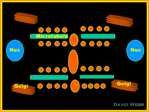
Phragmoplast (Microtubules &
Golgi Vesicles) forms at Late Anaphase-Telophase
Microtubules provide the orientation
for Golgi Vesicles which contain Cell Wall Precursors that form the Cell Plate.
Following Nuclear Division the Phragmoplast forms near the center of the former
Metaphase Plate. The Phragmoplast is composed of Microtubules & Golgi Vesicles. The
fused central Vesicles are called the Cell Plate.
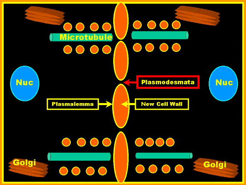
The vesicles from the two new cells move towards the center and fuse. The contents
of the Vesicles become the Middle Lamella of the new Cell Wall. The vesicles from the two
new cells move towards the center and fuse.The contents of the Vesicles become the Middle
Lamella of the new Cell Wall.
Microtubules appear to guide the movement of the Vesicles & these become more
peripheral as the new wall grows.There are gaps in the Cell Plate. These become
Plasmodesmata and connect the cytoplasm of both new cells. This process progresses from
the center towards the to the periphery until the New Wall joins the Old Wall.
Self Assembly Hypothesis
The various polymers which form the
wall (Cellulose - Hemicellulose - Pectins) have an inherent capacity to
self-organize and may not need complex biochemical process for this to occur.
Wall Enzymes -> Polymerization
Alternatively, there may be enzymes in the
Cell Wall which participate in the polymerization of these molecules.