 The larval brain forms during the 24 hours of embryogenesis. Subsequently, Drosophila
goes
through three larval stages (L1, L2 and L3) before it pupates and then reaches adulthood.
The larva has a number of specific brain structures that are replaced by different adult
structures during metamorphosis. The larval brain already contains the anlagen for the
adult brain. The visual system of the larva consists of Bolwig's organ, which is connected
to the larval optic neuropil via Bolwig's nerve. These three structures, for instance, are
degraded or retracted during metamorphosis. They are replaced by the adult visual system
with the four neuropil structures lamina, medulla, lobula and lobula plate. These start to
develop in larval stage L3 and continue their development during pupation.
The larval brain forms during the 24 hours of embryogenesis. Subsequently, Drosophila
goes
through three larval stages (L1, L2 and L3) before it pupates and then reaches adulthood.
The larva has a number of specific brain structures that are replaced by different adult
structures during metamorphosis. The larval brain already contains the anlagen for the
adult brain. The visual system of the larva consists of Bolwig's organ, which is connected
to the larval optic neuropil via Bolwig's nerve. These three structures, for instance, are
degraded or retracted during metamorphosis. They are replaced by the adult visual system
with the four neuropil structures lamina, medulla, lobula and lobula plate. These start to
develop in larval stage L3 and continue their development during pupation.
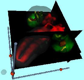
The larval brain reconstruction presented here consists of the following structures: Outlines of both brain hemispheres and the ventral ganglion (grey, transparent), central brain and ventral ganglion neuropil (red), inner optic anlagen (green), larval optic neuropils (purple), parts of Bolwig's nerves (blue), developing medullae (gold), and developing laminae (yellow). The surface reconstructions are a result of segmenting each optical section of a double-labeled larval brain by hand. An antibody staining against Synapsin shows all active neuropils (central brain and ventral ganglion, larval optic neuropils). Secondly, GFP driven by the enhancer trap line Mz1369 shows the developing visual system within the larval hemispheres. Both, surfaces and original can be analyzed in three dimensions using the reconstruction.