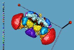
 The adult brain consists of neuropil, fiber tracts and a surrounding cell body layer. The
reconstruction only shows neuropil labeled by antibody nc82 and the surrounding cell
body layer (shown here in transparent green). The adult neuropil consists of a small
subesophageal ganglion (deep purple) and a large supraesophageal ganglion (other colors).
They are intimately fused which makes it difficult to find a clear demarcation between
them. In other insects, in which the head ganglia are separated, the subesophageal ganglion
is the relay station between the supraesophageal ganglion and the ventral cord. In flies
part of the traffic bypasses the subesophageal ganglion. The supraesophageal ganglion is
structured into characteristic neuropil regions, which can be identified in nearly all
insects. The most conspicuous ones are color-coded in the model and described below.
The adult brain consists of neuropil, fiber tracts and a surrounding cell body layer. The
reconstruction only shows neuropil labeled by antibody nc82 and the surrounding cell
body layer (shown here in transparent green). The adult neuropil consists of a small
subesophageal ganglion (deep purple) and a large supraesophageal ganglion (other colors).
They are intimately fused which makes it difficult to find a clear demarcation between
them. In other insects, in which the head ganglia are separated, the subesophageal ganglion
is the relay station between the supraesophageal ganglion and the ventral cord. In flies
part of the traffic bypasses the subesophageal ganglion. The supraesophageal ganglion is
structured into characteristic neuropil regions, which can be identified in nearly all
insects. The most conspicuous ones are color-coded in the model and described below. The optic lobes are located adjacent to the eyes and each consists of four neuropil regions, lamina (not shown), medulla (red), lobula (orange) and lobula plate (light orange). In this preparation the lamina has been removed together with the eye and head capsule. Each neuropil region consists of an orderly array of ca. 750 columns. These columns, the neuroommatidia, reflect the retinotopic organization of the optic lobe, i.e. fibers preserve their neighborhood relations throughout the ganglia and each neuroommatidium evaluates retinal inputs with the same point of view. Stacks of fiber strata perpendicular to the columns provide crosstalk between the visual elements. Large field tangential neurons connect the optic lobes of the two hemispheres and sets of projection neurons transmit the preprocessed visual data to the central brain.
The most prominent structures in the central brain are the antennal lobes (yellow), the mushroom bodies (brown) and the central complex. The antennal lobes (red) consist each of 40 identified glomeruli, which receive the olfactory receptor fibers of the 3rd antennal segment and the maxillary palp. The glomeruli collect fibers of the same chemosensitivity and relay their activity to projection neurons and antennal lobe intrinsic fibers. The latter neurons connect several or all glomeruli providing inhibitory or excitatory crosstalk between them, the former carry the preprocessed primary chemospecificities to the brain.
The mushroom bodies (brown) are a paired structure. The mushroom body neuropil is a tight bundle of about 2500 thin parallel fibers (Kenyon cells). The best known input neurons are projection neurons which connect single glomeruli of the antennal lobe to the lateral horn (pink) sending side branches into the posterior part of the mushroom body, the so-called calyx.
The central complex lies in the middle of the brain above the esophagus and just in front of the large commissure (pale pink), the main fiber tract linking the two brain hemispheres. It consists of four main regions, 1) the protocerebral bridge (yellow) at the dorso-caudal margin between the two calyces, 2) the fan-shaped body (dark green), an ordered array of columns, strata and shells, 3) the ellipsoid body (green), a perfect ring of neuropil, and 4) the noduli (light green), a small, compact lobe underlying the fan-shaped body. Input reaches the central complex primarily through large-field tangential neurons, the main output projects via columnar neurons to the ventral bodies, which in turn are directly connected via the cervical connective to the ventral ganglion.