|
||
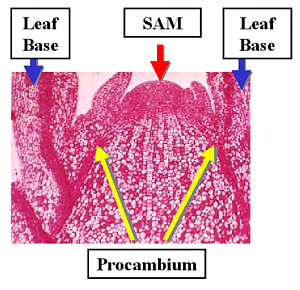 |
Procambium develops near the base of new leaves which originate from the Shoot Apical Meristem (SAM). We will follow the development of Xylem in the right-hand side of the image. | |
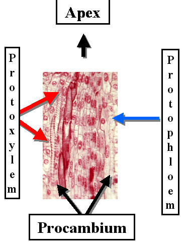
 Long Section through Procambium showing Protoxylem differentiation on the left side. |
Procambium consists of elongated cells which divide longitudinally near the shoot tip. When cells stop dividing they begin to differentiate. It is relatively easy to follow xylem differentiation because tracheary elements form characteristic secondary wall thickenings. Remember that the first xylem to differentiate is Protoxylem. Find the Protoxylem in the picture of Procambium on the left. | |
  |
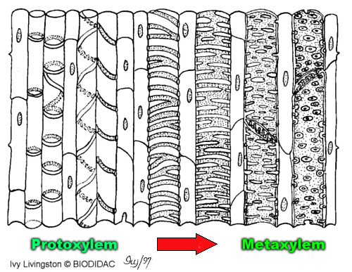
The first secondary walls are deposited as individual hoops which are NOT interconnected. These are called Annular Rings. This gives some lateral support but also allows the cells to stretch longitudinally. This is important because the growing point needs water to survive but its growth would be constrained by xylem cells with interconnected secondary walls. These cells resemble a reinforced hose on an old vacuum cleaner or the radiator hose of cars. Cells that differentiate later form Helical Thickenings. These create spirals like a Slinky. |
|
Protoxylem Cells with Annular Rings |
 Protoxylem Stained with Toluidine Blue Locate the Annular Rings & Spiral Thickenings! |
|
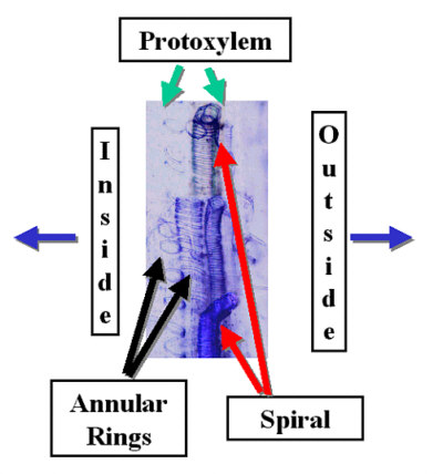 |
The next Tracheary Elements have spiral (helical) thickenings and resemble stretch socks that contain a slinky toy. This provides greater support but also allows cells to stretch and grow. Both of the above occur in the Protoxylem. Protoxylem forms in rapidly growing areas. However, it may be lost by the shearing forces of elongation or by compression due to lateral cell enlargement. |
|
|
|
More complex, interconnected wall patterns form once elongation has ceased. These xylem cells comprise the Metaxylem. They provide much greater support and are more permanent than Protoxylem. One of the most common wall thickening patterns is called Scalaraform. These walls have thickenings which resemble rungs of a ladder. The spaces between the rungs have only primary cell walls. |
|
 |
Tracheary elements which have most of their surface area covered by thick secondary walls, except for small openings are said to be pitted. The small openings are PITS . |
|
This Cartoon
Illustrates the |
||
|---|---|---|
| Syllabus | ||