|
 Prior Page |
 Next Page |
|
Prior Page |
Next Page |

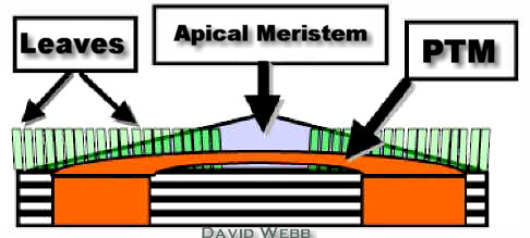
In many cases there are no differences between Monocot and Dicot Shoot Apical Meristems (SAMs). However some monocots like Coconut Palm, Banana and Ti (Cordyline) have a Meristem which subtends the SAM. Its meristematic cells divide anticlinally near the SAM, and periclinally as they are displaced from the SAM. This meristem produce files of cells towards the inside of the stem. This causes an increase in the diameter of the stem in the vicinity of the SAM with virtually no Internodal Elongation. This is called the Primary Thickening Meristem (PTM)and is only found in monocots. Procambium is abundant in the derivative cells of the PTM. These are the primary vascular bundles of the stem. These plants have many veins in their leaves and this is reflected in the number of vascular bundles in the stem.
Primary Thickening Meristem in Ti |
|
|
|
|
|
|
  |
| Primary Thickening Meristems in Monocots | |
Monocots like Sugarcane and Corn do not have primary thickening meristems but they do have numerous Vascular Bundles in their leaves and consequently in their stems. The distribution of the Vascular Bundles in monocot stems looks disorganized at first glance. However, they are very well organized, but their organization is complex compared to Dicots.

While Asparagus is not the plant usually presented in textbooks, it shows traits which are similar to those in Corn & Sugarcane.
|
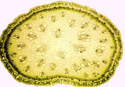
| Cross-Section of Asparagus Stem - Note the number and size diversity of the Vascular Bundles. |
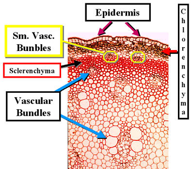
The stem is covered by an Epidermis which has a thick Cuticle & Stomata.
The outer part of the stem is photosynthetic and contains Chlorenchyma and small Vascular Bundles.
This is followed by a zone of thick-walled Sclerenchyma cells.
The rest of the stem is composed of Parenchyma in which Vascular Bundles are embedded.
There is no Pith as vascular bundles can be found near the center of the stem.
There is a progression in the size of the Vascular Bundles from small to large as one proceeds from the outside to the center of the stem.
|
|
|
|
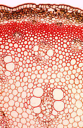
| Large Vascular Bundles in Asparagus | |
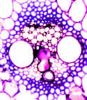
The vascular bundles (VBs) in corn (Zea mays) are "classical" Monocot VBs. They are surrounded by a Bundle Sheath which can be sclerotic. The Phloem has a highly organized appearance. The cells with the greatest diameter are the Sieve Tube Members. The smaller cells are Companion Cells.
The Xylem consists of large Metaxylem Vessel Members and smaller Protoxylem. Corn stems elongate rapidly and many of the protoxylem tracheary elements are destroyed. Consequently, the TEs may be missing and a protoxylem cavity (lacuna) can be seen instead.
|
|
| Vascular Bundles from Corn Stem - The Classic Monocot | |
|
Sugarcane Vascular Bundle Stained with Toluidine Blue Compare with the VB from Corn & Identify the same structures that are labeled above. |
|
Ancient Hawaiians used the stems of makaloa to make fine sleeping mats. These are considered to be the finest mats made by Polynesians. Some of these contained Pawehe designs. Mats with Pawehe were made only for royalty. The plants used to make the Pawehe designs were called kohekohe (Eleocharis calva). Both plants belong to the Cyperaceae (Sedge Family), and they grow in semiaquatic environments. We will study the stem anatomy of makaloa. Compare this stem with Asparagus.
The Stem is nearly circular in outline. Photosynthetic tissue is obvious in the outer region of the stem. Vascular bundles are visible at low magnification and they are distributed throughout the stem. There are also prominent air chambers throughout the stem.
|
Cross-Section of Makaloa Stem Unstained |
The presence of vascular bundles amid the photosynthetic parenchyma becomes obvious at higher magnification.
|
Cross-Section of Makaloa Stem Unstained |
The presence of fiber bundles just beneath the Epidermis becomes apparent at higher magnification.
|
Outer part of Makaloa Stem |
A thick cuticle is observed at higher magnification. The Chlorenchyma cells are distributed around the small, peripheral vascular bundles.
|
| Epidermis and outer region of Makaloa Stem, Unstained and stained with Toluidine Blue - Note the Cuticle, Fiber Bundles, Chlorenchyma & Small Circular Vascular Bundles. |
Staining with Toluidine Blue shows that most of the cell walls are lignified. It also makes the air cavities more obvious, see below.
|
Makaloa Stem Cross-Section stained with Toluidine Blue Note the Aerenchyma. |
|
Interior Vascular Bundles of Makaloa Stained with Toluidine Blue Note the Fibers which surround each VB as well as the location of Phloem & Xylem. |
Larger Vascular Bundles, similar to those in corn and sugarcane are observed towards the center of the stem. They are surrounded by Bundle Sheaths composed of thick, lignified Sclerenchyma.
Wheat is one of the most important crops in the world. Its cultivation is linked to the beginnings of civilization in the Middle-East which is sometimes called the cradle of civilization. The An Eagle is at the center of the Great Seal of the United States of America. He holds shafts of arrows in one talon & shafts of wheat in the other! Wheat cultivation played an important role in the development of the USA across the Great Plains.
|
| Cross-Sections of Wheat Stem Note that there are two size classes of Vascular Bundles. Also note the Photosynthetic Parenchyma. |
The center part of a mature wheat stem is hollow due to the rapid rate of stem elongation which occurs in stem development. The rim of tissue that remains contains vascular bundles of various sizes. The smaller bundles are associated with chlorenchyma in the outer stem. Larger bundles are seen towards the inside of the stem. Most of the cells may be sclerotic and stain positively for lignin. Stomata are present in the Epidermis. These are associated with the pockets of photosynthetic parenchyma.
|
Prior Page |
 Top |
Next Page |