|
 Prior Page |
 Next Page |
|
Prior Page |
Next Page |
We customarily analyze cross-sections as these give us the most anatomical detail and they are easy to comprehend because they are two-dimensional. However, it is important to understand how tissue systems are integrated within an organ like the stem. With the aid of computer programs we can hopefully illustrate this.
We will explore the most simple example. In this scenario,
Individual vascular bundles will run separately throughout the length of the stem.
These will produce Leaf Traces at regular intervals. A leaf trace is a vascular bundle which diverges from the axial bundles in the stem and enters a leaf.
Each Leaf Trace consists of one Vascular Bundle.
In order for the axial bundle to continue, it must have a way to compensate for the vascular tissue lost via the Leaf Trace. Otherwise it would terminate!
In some dicots the axial bundle forms 3 branch bundles at the Node.
The central bundle enters the leaf (Leaf Trace).
The other two unite above the node and reconstitute the axial bundle.
A Simple Model
One way to visualize this is to say that your body is the axial bundle below the node. Your shoulders are the node & your arms and neck are the three branch bundles.
Place your hands above your head so that the palms touch. This is like the exercise called jumping jacks.
NOW, lean your head so the it is parallel to the floor.
Your head and neck become the Leaf Trace.
Your arms are the lateral bundles which reconstitute the axial bundles above the Node.
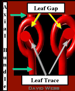 The cartoon above depicts two Vascular Bundles in a dicot stem. Start at the bottom of one axial bundle and proceed acropetaly. The axial bundles branch just below the node. One branch diverges into the leaf (Leaf Trace). The other two form arcs which converge above the node & reconstitute the Axial VB. Parenchyma occupies the area circumscribed by the curved lateral bundles. This looks like an area which has lost its VB when seen in cross-section. It is called the Leaf Gap. |
|
| Sectional view of a Leaf Gap - The bundle on the left has the same orientation as seen above. The bundle on the right is seen in a cross-section. Note the two small lateral bundles. These are aligned with the circumference comprised of Vascular Bundles in the stem. The area of parenchyma between them is the Leaf Gap. | 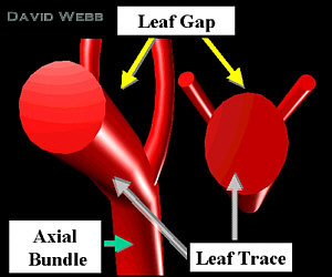 |
| By linking these together we get a simple Dicot Vascular System. | 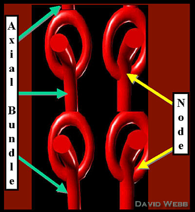 |
As you might guess, I am saving the best for last. Many familiar plants in Hawaii are monocots. This includes, Makaloa, Coconut Palm, other palms, hala, Sugarcane, Taro & Ginger. Consequently, it is important to understand the basics of monocot vascular organization!
Remember that there are many vascular bundles of different sizes in many monocot stems. Vascular bundles are highly branched in monocots and produce bundles of varying size and trajectory in the stem. These can change their trajectory and their size. This sounds hopeless but we are undaunted!!!! Aren't we??????
I will try to make a simple model which depicts the most important aspects of this.
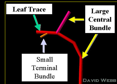 |
| The Large Central Vascular Bundles travels vertically over many nodes. However, their trajectory changes such that they diverge towards the base of a leaf. They produce a large branch which will resume growth as a large central bundle. They also produce small bundle(s). I have only included one which terminates near the Epidermis of the stem. The largest part of the divergent central bundle becomes a Leaf Trace. |
| As Jose Banderas said "Let's Play!!!!!!!!" |
 |
 |
 |
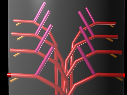
| This is how one half of a Vascular System might look in a stem. |
 |
| Make a mirror image & we have approximately half of the Vascular System. If we added mirror images towards the front & rear, we would have a complete system. However, if you understand this you can imagine the rest!!! |
 |
| Add
an Epidermis and Ground Tissue & we have a simple 3-D Model!!!! Relate this to the cross-section of Makaloa below! |
 |
|
 Prior Page |
 Top |
 Next Page |