 Phaeophyta - Brown Algae-6
 Phaeophyta - Brown Algae-6

Laminariales Anatomy
Three Distinct Tissue zones can be seen in many cases with the Laminae and Stipe.
These are the Medulla, Cortex and Meristoderm (Epidermis).
The central Medulla usually has loose filaments. However, parts of the Stipe Medulla have Parenchyma cells with thick cell walls. These support the Stipe.
Meristoderm & Cortex  Medulla |
 This is a composite image that shows the basic pattern of tissues in a Stipe. |
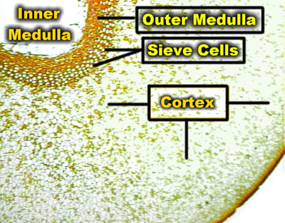 Cross Section of a Laminarian Stipe |
|
Phloem-like cells can be present in the Outer Medulla.
These have Sieve Plates & a type of Callose and are called Sieve Cells. The Sieve Cells in the Phaeophyta are found in the outer Medulla. They may also may have thick walls. Thick walls can be found in the Sieve Tube Members of flowering plants. Consequently, this is another trait that the Phaeophyta Sieve Cells share with Angiosperm Phloem.
Physiological studies have shown that sugars produced by Photosynthesis are conducted through these cells. Consequently, they are functional.
|
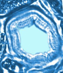 Sieve Cells cells may have relatively thick Cell Walls |
The Cortex of the Stipe has Parenchyma cells that have definite Pits with Plasmodesmata. They resemble parenchyma tissue in Angiosperms.
Large Secretory Ducts may be present.
A Meristoderm which resembles an Epidermis is also present.
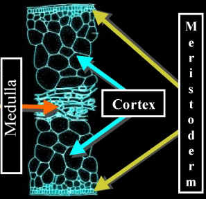
The Lamina has a similar organization with a central Medulla of loose filaments and a Cortex plus Meristoderm.
 Nereocystis Lamina |
 |
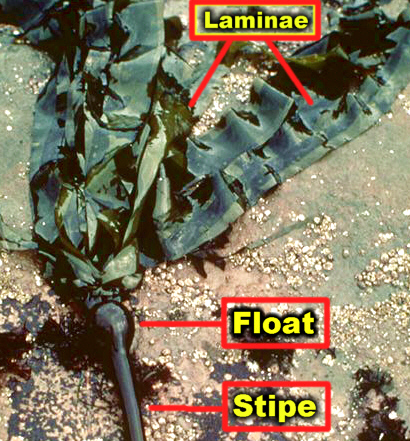
They help to keep Sargassum species afloat near the surface.

