Marchantia (Hepatophyta)
![]() Thallose Liverworts
Thallose Liverworts ![]()
Marchantia (Hepatophyta)
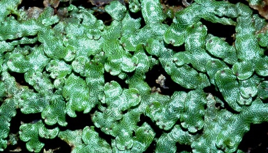
Marchantia Thalli: These have Apical Cells & Branch Dichotomously at the Apex. They are Dorsiventral in that they have distinct upper (Dorsal) and lower (Ventral) surfaces which comprise its Epidermis. They have a reflective Dorsal Epidermis which produces a distinct Cuticle. The white flecks on the surface of the Dorsal Epidermis are stomata-like Pores. |
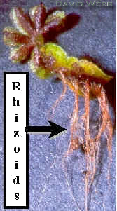
The Ventral Epidermis produces large Rhizoids which anchor the thallus to the substrate and may participate in water uptake. They grow in wet areas and can form dense mats. |
| Dorsal Epidermis of Marchantia. Note the compartmentalization of the Thallus. This modular construction protects the whole organism from local trauma. Each compartment has some level of autonomy. Each compartment has one large Pore. |
|
|
|
 |
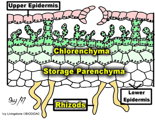 |
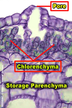
These sections through a Marchantia Thallus display the level of structural complexity achieved by this plant. We have already noted features of the Dorsal and Ventral Epidermis. A layer of Chlorenchyma is found directly below the Dorsal Epidermis. The photosynthetic cells are slightly columnar and there is a lot of air space between the cells. This is similar to the Mesophyll found in some "advanced" land plants. Note the way in which the Chlorenchyma distributed in relation to the Epidermal Pores. This is very precise and probably represents the most efficient distribution of Photosynthetic Cells and Pores. It would be interseting to look a Marchantia grown in different environments to see if this relationship varies with the environment. A layer of storage Parenchyma subtends the Chlorenchyma. The Ventral Epiderms produces Rhizoids which anchor the thallus and absorb water. Consequently, we see distinct layers which have different functions with regards to Photosynthesis and Water relations. |
|
![]()

