|
There has been a progression in the Complexity and Location of the Stele
This is largely related to the development of the Megaphyll which has large and numerous Leaf Traces that result in large Leaf Gaps.
The location of the Stele represents an equilibrium between the location of strengthening and conducting cells.
Engineering studies show that the ideal location for Strengthening Tissue is just beneath the surface of a cylindrical structure (Niklas 1997).
The ideal location for Conducting Tissues is the center of the axis where they would be least likely to break when stems are bent by wind.
It is instructive to note that Roots, which do not produce Leaves, have the most simple stelar types & the stele is in the center of the axis. In most cases this is an Actinostele. Furthermore the Stele is always at the center of the axis.
A painful review of the Steles in extant Land Plants
Bryophyta: The
"Stele" in the most complex Mosses is located in the center
of the stem. It produces minute leaf traces which connect the
Stele with the Leaf Vein "Nerve". There are no Leaf Gaps.
The Leaves are very small and simple.Strengthening tissues occur in the outer Cortex and/or in
the "Stele" (Sterids).
most complex Mosses is located in the center
of the stem. It produces minute leaf traces which connect the
Stele with the Leaf Vein "Nerve". There are no Leaf Gaps.
The Leaves are very small and simple.Strengthening tissues occur in the outer Cortex and/or in
the "Stele" (Sterids).
Psilophyta: The Stele is located
Lycophyta: The Stele Strengthening Tissues are still found in the
outer Cortex.
Pterophyta: The Stele complexity varies from a Haplostele to a Dictyostele to a Polystele. Megaphylls are present and more than one leaf trace may diverge to each Megaphyll. There can be many overlapping Leaf Gaps which result in a divided stele (Dictyostele). Dictyosteles are located in the Outer Cortex, rather than in the center of the stem. Rhizomes with no leaves tend to have simple, central Steles (Haplo, Actino, Plecto) Strengthening Tissue is primarily located in the Outer Cortex.
Anthophyta & Coniferophyta:
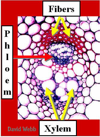
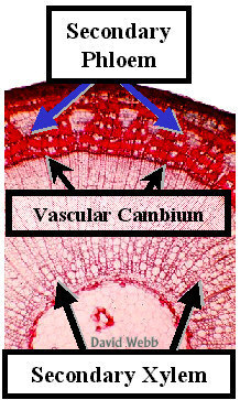
In Strengthening Tissue (Sclerenchyma) is associated with the Vascular Bundles, Leaf Traces and major Leaf Veins. Megaphylls rule and there are many Leaf Gaps in Dicot Steles. Monocot leaves have many major veins which are directly connected to a complex of highly branched vascular bundles in the stem. The Bottom Line is that increased Stelar Complexity and its more peripheral Location correlate with the presence of Megaphylls which have large, complex Leaf Traces & many Leaf Gaps. Strengthening Tissues are now associated with Vascular Bundles, Leaf Traces and principal Leaf Veins. The shift from a Eustele to Siphonostele is probably due to a functional shift for each stem segment as new ones are produced by the Shoot Apical Meristem & as Branches get larger. The Eustele & Atactostele are associated with soft stems that have actively photosynthesizing leaves. These are relatively light in weight and require little structural support. Furthermore, there is a need for more efficient radial transport out of and into vascular bundles of the leaf. Smaller Vascular Bundles are more efficient in terms of local radial transport than a large Stele located at the center of the stem, or minute Leaf Traces. The development of a Siphonostele from a Eustele occurs in parts of the stem which have shed their leaves & are far removed from the shoot apex. There is a greater need for structural support in these areas, due to the increased mass that accrues from Primary Growth (new Leaves & Stem tissues). There is also a greater need for Longitudinal Transport to and from the metabolically active parts of the stem tip that is getting further and further away from the Roots. Structural support is now provided by Secondary Xylem which is located near the periphery of the stem and extends to its center. Most of Xylem conduction occurs in its outer zone near the Vascular Cambium & Secondary Phloem.
|
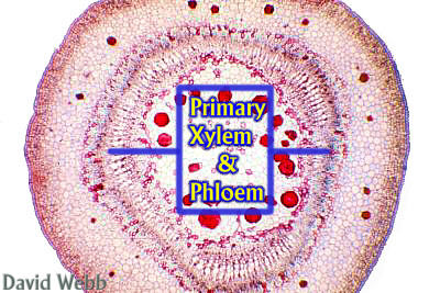
Cross-section
There is a Correlation between the type of Stele and |