Lycophyta-Selaginellales-3 |
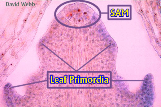
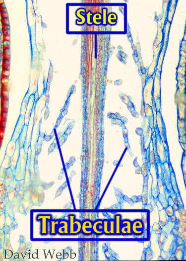
The Shoot Apical Meristem can be an Apical Cell OR it can be Multicellular.The Epidermis of the Stem has a cuticle but is otherwise unremarkable.The Stem Cortex contains Chlorenchyma, storage Parenchyma & Sclerenchyma.Leaf Traces are present. These are small Vascular Bundles that connect the leaf vein to the Stele in the stemThe Endodermis is unique and its cells are called Trabeculae. These cells are highly elongated and span an air space which intervenes between the Cortex and the Stele.They are also present in Leaf Traces.
The Function & Development of the Trabeculae are Unknown. Air chambers in plant organs are associated with Photosynthesis but the center of the stem is not photosynthetic.They are also associated with aquatic plants and probably store oxygen which is required for Respiration. They might also store CO2 because higher plants can't absorb bicarbonate which is the form CO2 takes in water. Water storage might also occur in the air space. Finally, this arrangement might allow the stem to be more flexible. Fascinating!!!!!!
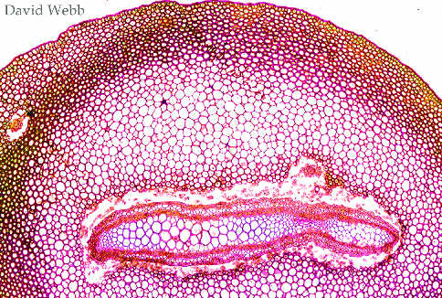
The Stele of Selaginella Stems is basically a Plectostele.However, there is often only one plate. Furthermore, the Plate may be Divided into separate bundles called Meristeles. Meri means to divide. Thus, Selaginella may have a divided stele.These appear to be separate but coalesce and are interconnected periodically along the axis of the stem. We will say that Selaginella has a Protostele.Roots arise at branch points along the stem. They have been called rhizophores because they don't look exactly like roots until they contact the soil. However, they are roots nonetheless. The root system is Adventitious! They branch dichotomously and have a Haplostele.The Vascular Tissues are similar to those of Lycopodium.
|
|
![]()