|
||
 |
Two unsuspecting Parenchyma cells on a stroll in the park. How are they to know that they will soon be transmogrified into Sclerenchyma cells. Note the overall cell shape and wall thickness. Nuclei are prominent in the thin peripheral cytoplasm. | |
 |
Sclereid under construction. Note the similarity in shape to the Parenchyma cell above. However, note the difference in wall thickness. The new wall is called Secondary and is deposited after the cell has ceased enlargement. The distinguishing features of Sclerenchyma are the presence of a Thick Secondary Wall which is highly organized, and usually contains Lignin. | |
 |
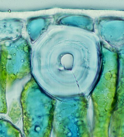
A large Sclereid. Note the laminated, thick secondary walls. The red stain indicates the presence of Lignin. There are two types of Sclerenchyma. Sclereids occur alone (idioblast) or in small clusters. They are usually isodiametric although some can be very long. They have prominent Pits & are generally lignified. | |
 |
These are clusters of Brachysclereids from pear flesh. | |
 |
The term Brachysclereid is used for isodiametric sclereids like this from pear. The secondary wall almost occludes the cell. It is traversed by large branching pits.The red color is due to Lignin. | |
 |
Large Sclereidfrom the leaf of Podocarpus. Note the wall laminations and the Pits around 3 & 5 o'clock. Viewed with polarized light. | |
Prior Page |
Home Page | Next Page |