|
||
 |
Collenchyma cells bear a strong resemblance to Parenchyma. However, they have some distinguishing traits. They occur in groups just beneath the Epidermis. They have a primary cell wall which contains lots of pectins. Thus, they stain pink with Toluidine Blue. The cell wall is unevenly thickened, however. | |
 |
The thickenings can occur at the corners of adjacent cells. This is called Angular Collenchyma. This is illustrated in this embossed image. | |
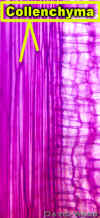 |
Longitudinal Section of Collenchyma which shows the Long Tapered shape of Collenchyma Cells. | |
Prior Page |
Home Page | Next Page |