|
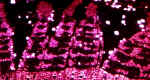
Longitudinal section of Pinus at the interface of secondary Xylem (Blue) and Secondary Phloem. |
|
|
The Vascular Cambium can be seen between the two Vascular Tissues |
|
|
|
|
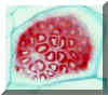
It is easy to distinguish Secondary Phloem and Xylem in a longitudinal section. The Xylem has stained positively for Lignin (Red). The Phloem has stained Blue. The freckled areas along the radial walls of the Sieve Cells contain Sieve Pores. Sieve Areas contain numerous Sieve Pores in small, discrete areas. Note the elongated shape of the Sieve Cells. |
 Sieve Areas |
|
Longitudinal Sections showing the darkly stained Parenchyma Cells & Sieve Areas |
 |
|
Longitudinal Section showing the darkly stained Parenchyma Cells. The light colored objects inside the Parenchyma cell on the right are amyloplasts. |
The Sieve Areas stain Blue in this preparation. |
|