|
|
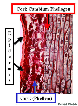
 The Epidermis has fallen away from this stem. As the diameter of the stem increases, the Epidermis cracks and eventually falls off. The Phellogen cells are usually the smallest ones in a file. Why is this so??? |
 |
| As the Diameter of the stem increases due to the activity of the Vascular Cambium, cracks would develop in the Periderm if only Periclinal Divisions occurred, as these produce radial files of cells without producing cells laterally. Consequently, Anticlinal divisions occur. These increase the number of cell files laterally and thus increase the circumference of the Phellogen & Phellem. | 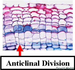 |
| We
are most familiar with the anatomy revealed by transverse or cross-sections. However, it
is important to envision anatomical features in longitudinal sections, as well. To the right you see a conveniently labeled long section through a stem in an early stage of Periderm formation. Relate this to the cross-sections above. |
 |