Lecture Directory |
Periderm
| As we know, there is a second
Lateral Meristem called the Cork Cambium. We need to focus our attention at the interface of the stem with the outside environment. |
|
| The Epidermis has stained blue which indicates the presence of Lignin and Suberin. Just beneath the Epidermis is a layer which is 4-5 cells thick. This constitutes a Periderm in an early stage of development. Cells in this tissue are organized in radial files. This indicates that they were formed by Periclinal divisions. If we trace the cell files back to their origin what would we find? |
|
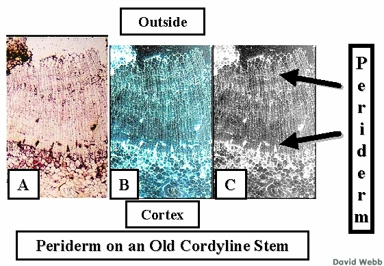 Periderm from an old Cordyline stem seen with color (A), Negative Color (B) and Gray Scale (C). Look for the long cell files which indicate the activity of a Phellogen (Cork Cambium). This is very similar to what we have seen with Dicots!!!!!! |
|
![]()


