Lecture Directory |
Lecture Directory |
| Meristematic
cells in the Primary Thickening Meristem do not stain as well as adjacent cells and appear
as a light band in microscopic preparations. Try to locate the radial cell files in the adjacent Photos. Concentrate on the far left region as indicated by one of the boxes. If you examine the stems of monocots that have secondary growth, you will see that the leaves are tightly clustered at the stem tip and that little internodal elongation occurs. You will see one leaf base on top of another, on top of another. See the image below. |
Closer View of the PTM in Banana. The |
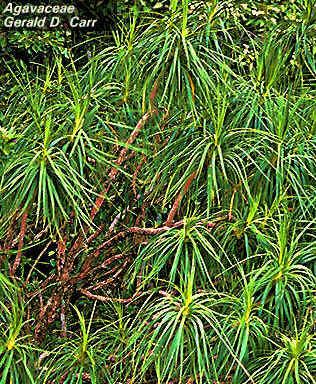
Hala pepe a local Monocot with Secondary
Growth
![]()


