VASCULAR CAMBIUM AND SECONDARY XYLEM
![]()
VASCULAR
CAMBIUM AND SECONDARY XYLEM
Vascular Cambium – Axial & Radial Systems
In
previous labs we studied the differentiation of primary xylem from the procambium,
and the beginning of secondary growth from the vascular cambium. We
used transverse sections to follow the production of secondary tissues. 
Transverse sections are cut perpendicular to the long axis of the stem and yield the most anatomical data. However, it is important to envision what happens parallel to the axis of the stem.
The term radial is applied to a longitudinal section which has a plane that is parallel to a radius that passes through the center of the stem.
Tangential sections are longitudinal but are cut perpendicular to the rays. We will say more about these later. Remember that transverse sections are cut perpendicular to the long axis of the stem.
Rays constitute the radial system in secondary xylem and phloem. As the name suggests rays run parallel to radii that pass through the center of the axis. They are composed of parenchyma cells which are usually elongated perpendicular to the long axis. Thus, we could call them radial (ray) parenchyma.
The term axial is applied to cells which are oriented parallel to the axis of elongation. Thus, we could study axial parenchyma. The axial vascular system is composed of xylem and phloem, plus parenchyma. Each of these contains axial parenchyma, specialized vascular cells and fibers. The radial system is simple by comparison. Occasionally, tracheids, laticifers and secretory ducts may be present in both systems (axial & radial).The axial
and radial systems can be studied with two types of longitudinal sections. 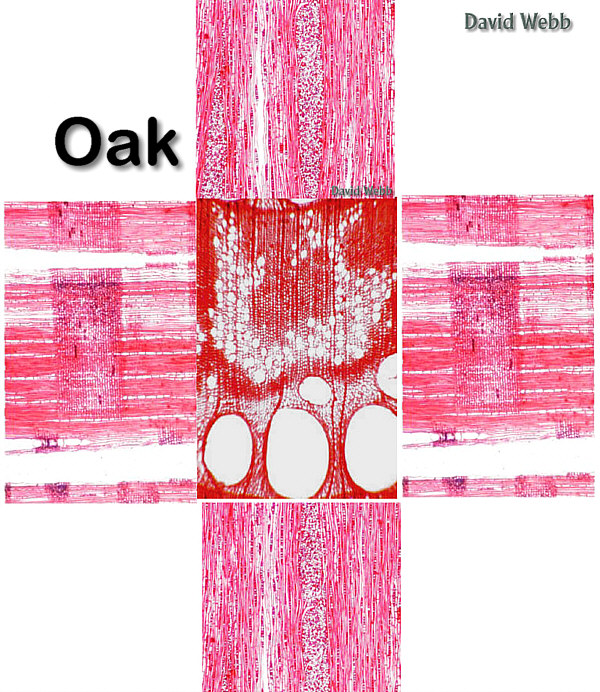
Radial sections are longitudinal sections cut parallel to the rays.
Tangential sections are cut perpendicular to the rays.
See if you can identify the planes of section for the cross-shaped image on the right.
In order to
refresh your  memory, examine transverse sections of Aristolochia
("D") slides. Transverse sections reveal the relationship of the cambial
cells to their immediate derivatives which become secondary xylem or phloem.
The young xylem and phloem cells form radial files because of their origin from the
tangentially dividing cambial" initials". In older xylem
memory, examine transverse sections of Aristolochia
("D") slides. Transverse sections reveal the relationship of the cambial
cells to their immediate derivatives which become secondary xylem or phloem.
The young xylem and phloem cells form radial files because of their origin from the
tangentially dividing cambial" initials". In older xylem  the radial seriation is disturbed because of non-uniform cell
enlargement. There is, for example, a striking difference in width between the vessels and
the cambial cells. The vessels greatly disturb the original radial file of cells produced
close to the cambial zone.
the radial seriation is disturbed because of non-uniform cell
enlargement. There is, for example, a striking difference in width between the vessels and
the cambial cells. The vessels greatly disturb the original radial file of cells produced
close to the cambial zone.
Secondary
phloem shows a more orderly arrangement because its cells do not enlarge greatly. The cambial
cells are relatively undifferentiated.
The phloem and xylem derivatives gradually acquire their characteristic traits. It is
instructive to follow the radial files of cells from the cambium into the mature tissues
and to recognize the differentiation processes that involve changes in size, wall
structure, and cells contents.
relatively undifferentiated.
The phloem and xylem derivatives gradually acquire their characteristic traits. It is
instructive to follow the radial files of cells from the cambium into the mature tissues
and to recognize the differentiation processes that involve changes in size, wall
structure, and cells contents.
 two
systems.
two
systems.
The ray initials are relatively small and occur in groups corresponding in width and height with the rays.
The initials of the axial system are elongated and pointed. They are usually called fusiform (spindle-shaped) initials. They are relatively short in Robinia. In tangential sections the fusiform cambial cells occur in horizontal tiers. Cambium thus organized is referred to as storied or stratified. The contrasting type would be non-storied or non-stratified cambium. In the non-storied cambium the fusiform initials overlap one another and do not appear in horizontal tiers in tangential views. Storied VC produces storied wood!!!!!!

Tangential Section of Storied Wood
Examine tangential sections of the vascular cambium of Juglans (Walnut). Is this storied or non-storied? What else can you determine by observing the vascular cambium?
Ring-porous
wood has wide vessels (pores) concentrated in the early wood and
smaller vessels in the late wood.

Diffuse-porous wood has vessels of relatively uniform size distributed evenly throughout growth intervals.

Variations in Wood Structure
Examine
blocks of wood from several plants and note the following:
a] growth rings,

Compare several kinds of wood, using thin sections.
Certain woods have conspicuously wide pores, the vessel members while other woods do not have vessels but have tracheids.
If vessels are present, they may be filled with tyloses. What are tyloses?
In some woods rays are only one cell wide (uniseriate). In others the rays are several cells wide (multiseriate) and visible to the naked eye.
 |
|
| Tangential Section of
storied wood which has Uniseriate & Multiseriate Rays |
|
Growth increments or rings vary in width. The variation is due to the interaction of genes and the environment.
The features listed above are, in combination, responsible for the pattern of wood "grain".
The
concept of axial and radial systems in wood
Observe commercial slides of pine wood which clearly illustrate the differences between the two. Note especially the difference in appearance of the rays in the three kinds of sections. Rays are the most useful structures for distinguishing between the radial and tangential sections.
 Radial Section |
 Transverse Section |
 Tangential Section |
This is a good
time to consult the world famous
Fold A Wood Consortium for Cosmic Consciousness!

Click on the button to bring up a full-sized image of Pinus wood.
Print it out & trim away the white areas.
Fold & secure with tape.
Identify the three planes of section.
Bordered Pits
Pine wood clearly reveals the complicated structure of conifer bordered pits. A Torus is present for conifer bordered pits but is absent in angiosperms.
A clear concept of the parts of the pits and their interrelation should be obtained by the study of pit-pairs in face views (radial sections) and in sectional views (transverse and tangential sections).

The Fence Analogy for Rays
I think of the rays as fences.
The radial section allows you to see the fences from the side. Thus you can see the arrangement of the different fence posts as well as the height of the fence. However, you can’t see its thickness.
The tangential view is like looking at the fence from one of its ends. You can see its height and thickness but you can’t see the sides of the fence or is length.
If you take a few steps to the right you can see the end of the fence and its side at an oblique angle.
 |
 |
 |
| Radial View of a Fence | Tangential view of a Fence | Oblique view of a Fence |
What would a Transverse view of a uniseriate or a multiseriate fence/ray resemble?
 Uniseriate |
 Multiseriate |
Pinus
a vessel-less gymnosperm.
Examine
slides of transverse, radial, and tangential sections, and macerated
wood.
The following questions may be used as a guide to your study.
What features cause the
difference in appearance between early wood and late wood?
What kinds of cells occur in the axial system?
What kinds of pits occur in these cells?
How are those pits distributed?
What is the size of the
rays in width and height?
What kinds of cells occur in the rays?
What kinds of pits occur in the ray cells?
What are the large
schizogenous intercellular spaces found in the axial and radial systems of the wood? The
parenchyma cells lining them may occlude these spaces. If you don't know this by now, you
haven't been paying attention!!!! Boy you sure know how to hurt a guy!!!! Just when I
was beginning to like this course!!!!!!
Hey!!! This is just like
the Fold A Wood!!!!!
Examine macerated pine wood. This will help you answer the questions above.
Quercus (oak)
Pine wood is extremely simple!
Oak wood is extremely complex!
Examine
The following questions may be used as a guide to your study.
What features cause the differences between early wood and late wood?
What kinds of cells occur in the axial system?
What kinds of pits are found on the various cells of the axial system?
Are the vessels all the same width?
How are the vessels
distributed in an annual ring?
What name is given to a wood that has a distribution of vessels like the oak wood?
Are the ends of the vessel members oblique or transverse?
What type of perforation
plate is found in the vessel members? Hint Hint!!
Don’t spend too much time looking.
What is
the developmental effect of the large vessels on the arrangement of their surrounding
cells ?
How is the xylem parenchyma distributed as seen in transverse sections?
What term is applied to xylem parenchyma distributed in this manner?
What are the cells with particularly thick walls and narrow lumina?
What types of pits occur in these cells?
What are the cells that are intermediate in width between the thick-walled cells and the vessels?
What kind(s) of rays
occur in this wood?
What kinds of cells occur in the rays?
What are the principal differences between pine wood and oak wood?
The CIA and DDT must have had a hand in developing these questions!!!
Examine prepared slides or freshly macerated material.
Identify the cell types present. Try especially to differentiate between vessel members, tracheids, and libriform fibers. Note also the "tails" on some vessel members. This feature indicates the occurrence of intrusive growth.
 |
 |
 |
| A Large Vessel Member with Tracheids and Fibers | An elongated Vessel Member; Note the simple Perforation Plate | A Vessel Member (Left) with a Fiber-Tracheid (Middle) and axial Parenchyma (Right) |
 |
 |
 |
A Tracheid: Note the shape and presence of many Pits. |
A Tracheid: Note the many Pits | Libriform Fibers: Note the shape and diameter. |
Planes of Section (Optional)
We are most familiar with transverse or cross sections. These are made by slicing a structure along a plane that forms a right angle with its long axis. In other words the plane of section is perpendicular with the long axis. Transverse sections generate the greatest amount of anatomical detail, and are sufficient for many types of comparisons. However, in order to get a three-dimensional perspective on wood anatomy, it is important to use longitudinal sections.
Obtain cylindrical sections cut from Podocarpus and Coffee (Coffea) stems.
Use a new Razor Blade to cut semi-thin Transverse, Tangential & Radial sections by looking at the Illustrations below. These do not need to be extremely thin to be useful.
Place these on microscope slides & flood them with Phloroglucinol.
Allow these to stain for 5 – 10 minutes. (Don’t let them dry out!)
Add a coverslip & place the slide onto the microscope stage.
Use the 4X Objective but swing in the High Power Condenser Lens.
Turn the illumination to Maximum & look for the Rays in the Secondary Xylem.
What characteristics can you see for these rays? What is their height in terms of the number of cells which comprise them? Are they uniseriate or multiseriate? homocellular or heterocellular?
Also look for cells that comprise the Axial System.
Follow the xylem cell files back to the Vascular Cambium. How will you know where the xylem ends and the Vascular Cambium begins?
Fold A Wood
Cut out the cross shaped wood images below and fold them to make a cube.
Identify the different Planes of Section!
|
 |
|
SECONDARY XYLEM.
In the
previous labs we have looked at the origin and development  of
primary and secondary vascular tissues. We will examine details of secondary xylem for
some "typical" angiosperms and gymnosperms.
of
primary and secondary vascular tissues. We will examine details of secondary xylem for
some "typical" angiosperms and gymnosperms.
Tilia americana (basswood).
Examine prepared slides of transverse, radial, and tangential sections. Pay particular attention to the differences between oak and basswood.
 |
 |
 |
Name The Plane of Section
The following questions may be used as a guide to your study.
Yes Drill Sargent!!!!!
What features cause the difference in appearance between early wood and late wood?
What kinds of cells occur in the axial system?
What kinds of pits are found on the various cells of the vertical system?
Are the vessels all the same width?How are the vessels distributed in an annual ring?
What name is given to a wood that has a distribution of vessels like the Tilia?
Are the ends of the vessel members oblique or transverse?
What type of perforation plate is found in the vessel members?
Nonfunctioning vessels may appear to have partitions in them. What is the explanation of this phenomenon?
What is the developmental effect of the large vessels upon the arrangement of the surrounding cells of the vertical system and the rays?
How is the xylem parenchyma distributed as seen in transverse sections?
What term is applied to xylem parenchyma distributed in this manner?
What are the cells with particularly thick walls and narrow lumina?
What types of pits occur in these cells?
What are the cells that are intermediate in width between the thick-walled cells and the vessels?
What kind(s) of rays occur in this wood?
What kinds of cells occur in the rays?
What are the principal differences between the basswood and the oak wood?
Pseudowintera colorata.
Study prepared slides of transverse, radial, and tangential sections and macerated wood.
 Transverse Section |
 Radial Section |
The questions below may be used as a guide to your study.
Oh NO!! I can't take this!!!!!
What kinds of cells occur in the vertical system? (Note the absence of xylem parenchyma in the vertical system).
What kinds of pits occur in the cells of the vertical system?
Two very distinct kinds of rays are present. Describe them.
Can you see any indication of growth rings? If so, describe the changes in structure associated with such rings.
Compare the wood of a vessel-less Angiosperm (Pseudowintera) with that of a vessel-less gymnosperm (Pinus) in respect to the following features: a) types of rays b) types of pitting on tracheids.


Name The Plane of Section
Xylem Specialization (Time Permitting)
Two groups of prepared slides are provided. The slides in group A represent woods in which many primitive characteristics of the xylem are present.
In group B many specialized characteristics are exhibited.
Compare one or more species from each group noting such features as:
orientation of end walls;
types of perforation plates;
pitting on lateral walls of vessel members;
distribution of parenchyma;
types of rays.
It will probably not be possible to estimate lengths of vessel members in sections with any degree of accuracy but try to determine relative lengths of these cells in the two groups.
![]()
