|
|
This is a large and familiar taxon. We are
only studying the  Filicidae.
However, there are several others that we are not considering. The Filicidae is the
largest and most wide-spread taxon and thus best represents this Division. Ferns do not
generally dominate ecosystems but they can be important in certain environments.
Tree ferns may be among the dominant species is
certain areas. This is especially true for the Hawaiian islands.
Ferns have greater ecological significance in Hawaii than elsewhere due to lack of
competitors which would normally displace them as sites mature. In some cases ferns are
the only significant ground cover in Hawaiian forests and open slopes. They are
consequently important for soil stabilization and erosion control.
Filicidae.
However, there are several others that we are not considering. The Filicidae is the
largest and most wide-spread taxon and thus best represents this Division. Ferns do not
generally dominate ecosystems but they can be important in certain environments.
Tree ferns may be among the dominant species is
certain areas. This is especially true for the Hawaiian islands.
Ferns have greater ecological significance in Hawaii than elsewhere due to lack of
competitors which would normally displace them as sites mature. In some cases ferns are
the only significant ground cover in Hawaiian forests and open slopes. They are
consequently important for soil stabilization and erosion control.
The class that we are studying
is characterized by pinnate leaf organization. This is evident
in their venation and the overall appearance  of their lamina. Another characteristic trait is Circinate
Vernation. Immature leaves are tightly coiled and tightly packed at the
shoot apex. The leaves begin to uncoil when favorable conditions occur. They resemble the
tops of violins and have been called "fiddle heads". They also resemble the
shepherd's crook and have been so named. The fiddle heads mature from the base towards the
tip (acropetal maturation). Consequently, the lower sections
of the petiole and blade mature first. The leaflets of
compound leaves also display Circinate Vernation.
of their lamina. Another characteristic trait is Circinate
Vernation. Immature leaves are tightly coiled and tightly packed at the
shoot apex. The leaves begin to uncoil when favorable conditions occur. They resemble the
tops of violins and have been called "fiddle heads". They also resemble the
shepherd's crook and have been so named. The fiddle heads mature from the base towards the
tip (acropetal maturation). Consequently, the lower sections
of the petiole and blade mature first. The leaflets of
compound leaves also display Circinate Vernation. 
Ferns have Megaphylls (large leaves). However, size is not the chief characteristic of Megaphylls. A megaphyll is a leaf which contains more than one vein and its leaf trace is associated with a leaf gap in the stele. A leaf gap occurs when the departure of a leaf trace from the stele results in the development of parenchyma rather than vascular tissues in the stele, just above the leaf trace. This gives the stele a dissected appearance like the Dictyostele on the right. Some ferns have very simple leaf organization with little mesophyll differentiation. Others have a complex anatomy which rivals the structural specialization of angiosperm leaves.
The concept of a leaf gap is a little challenging because you need to think in three dimensions. I am including some drawings by Salvador Webi who is my favorite surrealist artist.
A Leaf Gap is present when a Leaf Trace diverges from the stele without its immediate replacement by vascular tissue. I have tried to illustrate this below. The stele is a Siphonostele. Note that the Stele (Dark Green) forms a complete hollow cylinder below the node. The cylinder is broken into two crescent-shaped columns near the Node because some of the vascular tissue appears to have diverged from the stele. The divergent vascular tissue is the Leaf Trace. The leaf trace enters the base of the Petiole and forms the midrib of the leaf. The gap in the stele contains parenchyma cells rather than vascular tissues. This area is the Leaf Gap.
The two drawings below attempt to show the anatomy of leaf gaps from a 3-D Perspective.
|
|
Three Dimensional Model of a siphonostele with leaf gaps. The leaf trace is emerging towards you & you are sitting on the leaf petiole. The stele is re-unified above the node and appears as a complete cylinder as seen in cross section. |
 |
 |
|
|
The presence of many overlapping leaf gaps produces a highly dissected stele, like a Dictyostele or a Eustele.
 Stems
StemsThe stems are usually subterranean. These can be a Caudex (erect) or a Rhizome (horizontal). Some species have Stolons (above ground & horizontal) or Erect Aerial Stems. Tree ferns are an example of the latter.
Branching is
sporadic and may be dichotomous or lateral.
There is little internodal elongation in most  species,
however there are viney types which are exceptions. Careful examination of tree fern
stem will show that it is composed of leaf bases which are contiguous
because there is No Internodal Elongation. This is an obvious
limitation in an environment that contains angiosperms. The latter generally have lots of
internodal elongation, and can quickly form a dominant canopy over slow-growing plants.
The leaves are tightly packed close the the shoot apical meristem which is an Apical
Cell and its most recent derivatives.
species,
however there are viney types which are exceptions. Careful examination of tree fern
stem will show that it is composed of leaf bases which are contiguous
because there is No Internodal Elongation. This is an obvious
limitation in an environment that contains angiosperms. The latter generally have lots of
internodal elongation, and can quickly form a dominant canopy over slow-growing plants.
The leaves are tightly packed close the the shoot apical meristem which is an Apical
Cell and its most recent derivatives.
The Filicales are Homosporous.
The spores are produced by sporangia located on the abaxial (lower) surface of the leaf.
The sporangia have a unique developmental ontogeny  (developmental sequence). They are Leptosporangiate
rather than Eusporangiate. Leptosporangia develop from a single
epidermal cell rather than a group of cells. The single cell produces an apical
cell. After a period of apical growth, Sporogenous tissue
differentiates and is surrounded by two - three layers of nonsporogenous
cells. The outer layer has an Annulus which contracts as the
sporangium matures such that the wall ruptures at a weak spot (lip), thus releasing the
spores. The annulus is sensitive to humidity and flails about
(developmental sequence). They are Leptosporangiate
rather than Eusporangiate. Leptosporangia develop from a single
epidermal cell rather than a group of cells. The single cell produces an apical
cell. After a period of apical growth, Sporogenous tissue
differentiates and is surrounded by two - three layers of nonsporogenous
cells. The outer layer has an Annulus which contracts as the
sporangium matures such that the wall ruptures at a weak spot (lip), thus releasing the
spores. The annulus is sensitive to humidity and flails about like a catapult. This helps to disperse the
spores. Sporangia may cover the entire abaxial surface
or large areas thereof. Sporangia may be limited to the leaf margin or veins, or may
occur in small clusters known as Sori
(heap or mound). The sporangia may be protected by a curled leaf margin or by an Indusium
(underwear). The indusium is a structure produced by the abaxial epidermis in association
with sporangia. It may be a simple flap of tissue or can be complex like an umbrella.
Indusia shield the developing sporangia. They are useful for identification.
like a catapult. This helps to disperse the
spores. Sporangia may cover the entire abaxial surface
or large areas thereof. Sporangia may be limited to the leaf margin or veins, or may
occur in small clusters known as Sori
(heap or mound). The sporangia may be protected by a curled leaf margin or by an Indusium
(underwear). The indusium is a structure produced by the abaxial epidermis in association
with sporangia. It may be a simple flap of tissue or can be complex like an umbrella.
Indusia shield the developing sporangia. They are useful for identification.
The Gametophytes
are usually photosynthetic and require light. They are typically bisexual but the
gametangia are separated in time, and by position. The Antheridia  form
first and are located near the posterior of the
gametophyte. The Archegonia are
produced later an are close to the apex of
the gametophyte.
form
first and are located near the posterior of the
gametophyte. The Archegonia are
produced later an are close to the apex of
the gametophyte.
This is not the first case in which the Antheridia matured first on Gametophytes. What if any significance does this suggest regarding sexual reproduction?
Embryos have a unique
development which is called Prone. The first
division of the zygote is longitudinal
rather than transverse. The embryonic shoot-root axis develops from lateral cells in the young
sporophyte rather than from an Epibasal or Hypobasal cell. There is no
dormancy and the sporophytes grow directly from the gametophyte.
axis develops from lateral cells in the young
sporophyte rather than from an Epibasal or Hypobasal cell. There is no
dormancy and the sporophytes grow directly from the gametophyte.
Leptosporangiate development and prone embryo development only occur in these plants. Consequently, the Filicidae comprise a divergent evolutionary line which does not lead to the Angiosperms (Flowering Plants).
These are beautiful organisms and have many horticultural uses.
We will analyze Laua'e (Phymatosorus scolopendrium) & a Sword Fern (Nephrolepis sp.) Kupukupu in detail.
You will also need to look at other specimens for individual features.
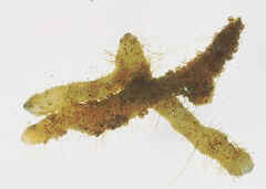
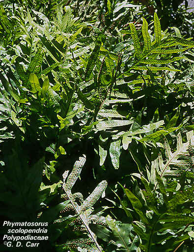 Laua'e Leaves |
 Laua'e Sori |
 Nephrolepis Leaves |
 Nephrolepis Sori - Note the Indusium |
Examine the Stolon or Rhizome or intact plants and note the origin of leaves, branches and roots.
Locate the position of the shoot apical meristem.
Does internodal elongation occur?
How would you describe the branching?
Is there evidence of Circinate Vernation? (Of course there is !!!!)
Are Scales present?
Examine the Leaves for the presence of Sporangia.
What term describes their clustered organization?
Can you find an Indusium?
Remove Sporangia with a dissecting needle and mount them in water on a microscope slide. Locate the Annulus and the Spores.
Mount some on a dry slide, place this under a dissecting scope or low power of your compound microscopes and breathe gently on them. What do you observe?

Study cross sections of the leaves, stems and roots prepared by us.
Note the key anatomical features.
Epidermal adaptations
Specialized Ground Tissues
Organization of the Stele
Examine other live plants for specified adaptations.
Examine these to gain insight regarding the organization of tissues in leaves, stems and roots.
Study Leaves of Cyrtomium with Sori.
Note the Anatomy of the Leaf and the Sori.
Is an Indusium present?
Can you observe Palisade and Spongy Mesophyll?
Why is this a Megaphyll?
Observe DEMO of Cyrtomium Sporangia
Examine Cross sections of other Fern Leaves & note the
Levels of complexity observed in the Mesophyll
Variety of Indusia
Examine slides of Polypodium Stem or Rhizome
Note the organization of the Vascular Bundles &
Identify the Type of Stele
Examine other Stems & Rhizomes to see the various types of vascular organization.
How does vascular complexity in Ferns differ from Psilotum & Lycopodium.
How does the complexity of fern leaves compare with leaves from Psilotum and Lycopodium?
Do you think these two are interrelated?
You should be able to Recognize Steles in a matching format.
Examine the DEMOs of
Pteris Apical Cell &
Pteris Shoot Apices (These are cut at various angles and are a little challenging)
Examine slides of Pteris Roots
What type of Stele occurs?
Where do branch roots originate?
Examine DEMO of Pteris Xylem (Longitudinal Section)
Note the Tracheids and the way in which they connect.
Examine Gametophytes, locate Gametangia
Observe Living Gametophytes
Examine commercial slides with Antheridia
Examine DEMOs of Archegonia
Examine DEMOs which show Sporophytes attached to Gametophytes. Connect the origin of the sporophyte with the gametophyte.
Which is the Dominant generation?
![]()