Myxomycota
Division: Myxomycota
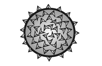
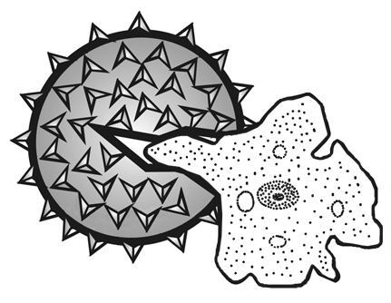
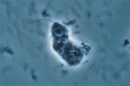
Members of this division are commonly referred to as slime molds. Although presently classified as Protozoans, in the Kingdom Protista, slime molds were once thought to be fungi (=kingdom Mycetae) because they produce spores that are borne in sporangia, a characteristic Myxomycota common to some taxa of fungi. However, the assimilative stage in slime molds is morphologically similar to that of an amoeba. This assimilative stage has been designated a myxamoeba (Fig 1). The myxamoeba, as is the case of the amoeba, is a uninucleate, haploid cell which is not enclosed in a rigid cell wall, and ingests its food by means of phagocytosis. During this mode of ingestion, the food particles, usually bacteria, beceome surrounded by the pseudopodia of the myxamoeba. Once the food has been engulfed in this matter, it is surrounded by a membrane or food vacuole where hydrolytic enzymes are secreted that will digest the food. In fungi, the assimilative stages are mycelium and yeast, both of which are surrounded by a rigid cell wall and obtain their food by means of absorption. These are some of the reasons why mycologists no longer recognize slime molds as being fungi. However, organisms in this group continue to be studied in mycology as a matter of tradition and not because they are thought to be related to fungi. Within the Myxomycota, the class Myxomycetes, often referred to as the acellular slime molds, will be the only class that we will consider.
Class: Myxomycetes
There are approximately 500 species of Myxomycetes. They are found on moist soil, decaying wood, and dung. One of the more interesting characteristic about this group of organisms is that while the species of other organisms will vary in different geographical localities, i.e. you don't find the same species of plants and animals on the mainland that you find in Hawai‘i, this is not generally true in the Myxomycetes. Most species can be found throughout the world.
Life Cycle of Myxomycetes
Introduction
Physarum polycephalum and Didymium iridis will be used to represent the Myxomycete life cycle and when possible, pictures of these species will be used to illustrate the various parts of the life cycle. These two species were selected since a great deal of researach has been carried out with these species and their life cycles are well known. However, since other species may differ somewhat in their life cycles, some of these variations will also be described.
Spore Germination, Myxamoebae and Swarm Cells
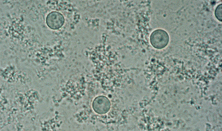
The spores of Myxomycetes are normally globose, uninucelate and haploid. The spore surface may range from almost smooth to reticulate. Spores of P. polycephalum and D. iridis are spiny. The spore wall is composed primarily of cellulose and is only one of two stages where a cell wall is formed. The other stage that forms a cell wall is the microcyst, which is discussed below. Upon germination, the spore will crack open and release a single, uninucelate myxamoeba. The myxamoeba moves by amoeboid motion and ingest food, by phagocytosis, as it does so. As the myxamoebae feeds and grows, they will reproduce, asexually, by mitosis and cytokinesis.
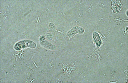
The myxamoeba stage may continue to proliferate for an indefinite period of time if there is available nutrient and the environment remains favorable. In most species, the myxamoeba stage may also vary according to the environment. When free water is available myxamoeba can differentiate into flagellated swarm cells (Fig 4-5). Although two flagella are present, One the long, anteriorly directed flagellum is visible. The second, very short flagellum is usually not visible. During periods of unfavorable conditions, the protoplast of the myxamoeba or swarm cell can round up and form a thin, cellulose protecetive layer around itself, called the microcyst (Fig 6), which will protect it from the environment.
Zygote and Plasmodium Formation
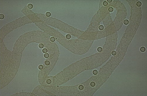
After a period of time, when a critical number of swarm cells or myxamoebae are formed, sexual reproduction will occur and these vegetative stages will then function as gametes. In P. polycephalum, it is the swarm cells that normally act as gametes. Gametes that are all derived from a common myxamoeba, however, are usually self sterile. In order for syngamy to occur, gametes that are derived from a different population of myxamoebae, and of a different mating strain, are required before syngamy can take place. By convention, the different mating strains are designated as a1, a2, a3, etc. and in order for syngamy to occur, the myxamoebae must be of different mating strains, e.g., a1 X a2, a1 X a3, etc. Once compatible mating strains have come into contact with one another, syngamy will occur to form the zygote. The zygote then undergo numerous mitotic divisions to form the large, multinucleate plasmodium. This class is commonly referred to as the acellular slime molds because the plasmodium (Figs. 7-8) stage of the lifecycle is not composed of many cells. Instead it is essentially a single, multinucleate cell. As in the myxamoebae stage, the plasmodium is also an assimilative stage that consumes food by phagocytosis. However, the plasmodium is a diploid structure and is many magnitudes larger. The appearance of the plasmodium is variable. In P. polycephalum (Fig. 7), it is a bright yellow, slimy structure while in D. iridis (Fig 8), the plasmodium is colorless. When unfavorable conditions prevail, the plasmodium forms a protective, brittle layer and becomes dormant. This dormant stage is termed a sclerotium (Fig. 9), and if observed under the microscope, it can be observed to be composed of a number of smaller multinucleate cells called macrocysts (Fig 10). Upon return of favorable conditions, each macrocyst can give rise to a new plasmodium.
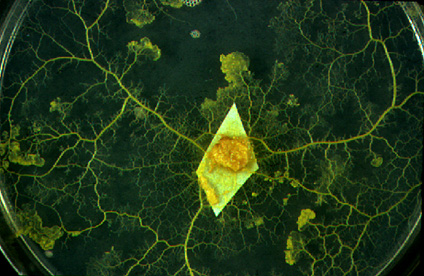 |
Figure 7: Plasmodium (2N) of Physarum polycephalum. A species with a bright yellow plasmodium. The plasmodium resulted from syngamy |
| of two compatible myxamoebae, followed by numerous mitotic divisions. | |
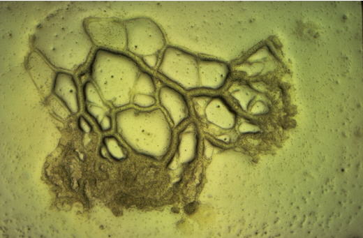 |
Figure 8: Plasmodium (2N) of Dydimium iridis. A species with a colorless plasmodium. |
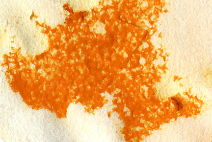 |
Figure 9: When conditions become unfavorable, a plasmodium can become dormant, as it did on this piece of filter paper, forming a resistant stage |
| that is a darker, yellowish-orange color called a sclerotium. | |
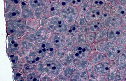 |
Figure 10: A section through a sclerotium shows that it is actually composed of smaller units called macrocysts. The number of nuclei in each |
| macrocyst are variable. | |
Formation of Sporangia
During favorable conditions, the plasmodium will migrate and feed for a period of time before being converted to numerous sporangia. In P. polycephalum, it is believed that the exhaustion of food is one of the stimuli that induces formation of sporangia because as long as adequate food is present in this species, the plasmodium stage will persist. Light appears to be another stimulus to fruiting in this species.
The form and color of sporangia are variable. In D. iridis, a light blue, more or less globose sporangium is produced on a yellowish stipe (Fig. 11) while in P. polycephalum the sporangium is a dark gray to almost black, lobed sporangium that is produced on a yellowish stipe (Fig. 12). The fragile, outer layer of the sporangium is the peridium (pl.=peridia), which may be persistent or degenerate by the time the sporangium is ready to disperse its spores.
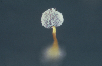 |
Figure 9: A single Dydmium iridis sporangium. The light blue powdery appearance of sporangium is due to the calcium carbonate crystal on the peridium surface. |
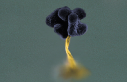 |
Figure 10: A single Physarum polycephalum sporangium. Note the numerous lobes in this species. Calcium carbonate is also present on the peridium |
| surface of this species, but is not obvious. | |
When sporangium formation begins, the plasmodium becomes denser and forms a thick sheet called the hypothallus (Fig. 11). The protoplasm of the plasmodium then becomes knotted into discrete nodules (Fig. 12), which represent the sporangial primordia. The individual nodules initially elongates (Fig. 13) and as development continues the basal portion, which will become the stalk, decreases in diameter while the upper portion becomes the sporangium proper and will develop the finger-like projection characteristic of P. polycephalum (Fig.14). In D. iridis, the sporangium would become a single, globose sac.
Upon the completion of movement of protoplasm into the sporangium proper, the stalk has now become more constricted and is devoid of protoplasm. Spore formation comes about with the formation of cell walls around the diploid nuclei. The nucleus in each spore will undergo meiosis to produce four 1N nuclei. Of these, three will degenerate. Thus, only one myxamoebae results from each spore. Also throughout the interior of the sporangium is the branched, thread-like capillitium. The capillitium arises from coalescene of vacuoles, which contain various material from the protplasm, especially calcium cabonate (CaCO3), in P. polycephalum and related species.
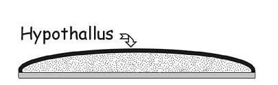 |
Figure. 11: A cross section through a strand of plasmodium with a thick hypothallus prior to sporangial |
| formation. | |
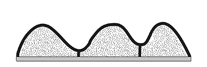 |
Figure 12: Cross section strand of plasmodium showing knotted, discrete units, with protoplasm pushing upwards. |
| Each unit will give rise to a sporangium. | |
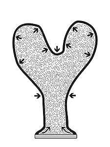 |
Figure 13: Longitudinal section of a single, developing, P. polycephalum sporangium. As the nodule elongates, the basal portion, which will develop into the stalk, becomes more and more constricted while the upper portion, which is the sporangium proper, continues to expand and forms the finger-like projections characteristic of this species. Arrows indicate points of constriction and expansion. |
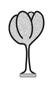 |
Figure 14: Upon completion of movement of protoplasm into the sporangium proper, the characteristic shape of P. polycephalum will have taken form.The stalk by this time will have become more constricted and devoid of protoplasm. Within the sporangium, following meiosis, the formation of haploid spores and capillium will occur. |
Sporangium Variations in Myxomycetes
The typical sporangia for P. polycephalum and D. iridis has previously illustrated. However, there are variations in sporangial types and structures. A few of these are illustrated below.
 |
Diachea leucopodia: Another species with sporangia that have stipes. Peridia are breaking apart in this picture to reveal the filamentous capillitium, beneath. |
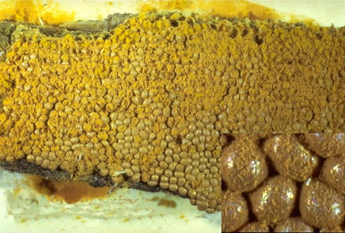 |
Trichia favoginea: Example of a species that produces sessile sporangia. Inset shows a close-up of tightly clustered sporangia. |
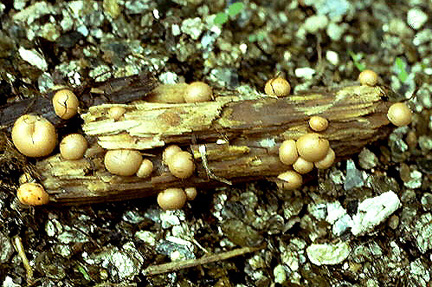 |
Lycogala epidendrum: Example of a species that produces aethalia. An aethalium resembles a sessile sporangium but is much larger. The larger size is |
| thought to have evolved from many smaller sporangia that have fused. | |
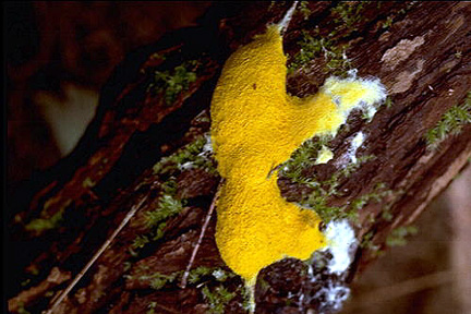 |
Fuligo septica: Another example of a species that produces aethalia. This species produces the largest known aethalium. |
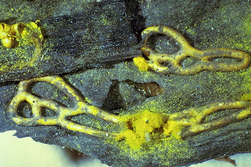 |
Hemitrichia serpula: Example of a species that produces plasmodiocarps. The spores and capillitium of this sporangium type retains the shape of the |
| plasmodial stage. | |
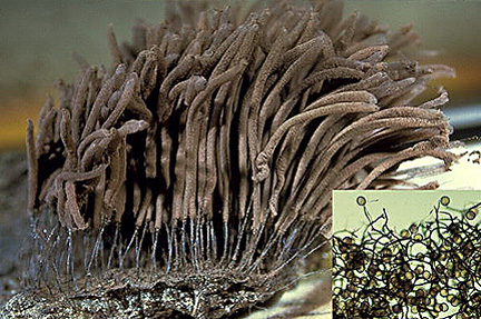 |
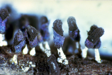
Stemonitis sp.: Example of a stipitate sporangium in which the peridium disintegrates at maturity thereby leaving the capillitium and spores exposed. Inset at |
| lower right shows capillitium and spores at high magnification. This species also differs in that stipe deveopment continues within the sporangium. The extension of the stipe is the columella. | |
Variations in Capillitium Morphology
Capillitium (pl.=capillitia) are filamentous structures that usually develop with spores within sporangia. They are thought to function in the retention of spores in the sporangia. By retaining spores the capillitium will allow gradual dispersal of spores over a long period of time. Thus, if over a period of time there are occasions when dispersal of spores will be unfavorable for myxamoebal and plasmodial growth, some spores will always be retained that will be dispersed at a later, possibly more favorable period. Capillitia are often ornamented and have been used in defining some taxa in Myxomycetes. Some variations are illustrated below:
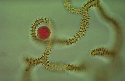 |
Trichia favoginea: Capillitium and spore (spore stained in 1% Phloxine). Capillitium ornamented with annular rings. |
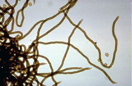 |
Hemitrichia serpula: Capillitium unbranched. Composed of strands of elaters. Elaters ornamented with double spirals, each coming from opposite |
| directions. | |
 |
Lycogala epidendrum: Capillitium (actually pseudocapillitium) without any distinctive ornamentation and spores. |