Division: Chytridiomycota
Members of the division Chytridiomycota have unicellular to mycelial thalli. Their cell wall composition is mostly chitin, and cellulose is not known to occur. Because cell wall composition is thought to be a conservative characteristic, this division was classified by Bartnicki-Garcia (1970) with the true fungi. However, it was not until studies sequencing of the small subunit rDNA was carried out that mycologist became bold enough to classify this division with the true fungi. Thus, this division has been classified with the true fungi even though flagellated spores and gametes are produced. Gametes and zoospores have a single, posterior whiplash flagellum. Sexual reproduction is variable and may be isogamous, anisogamous or oogamous. The division has a single class, the Chytridiomycetes, We will look at representatives from two orders, the Chytridiales and Blastocladiales.
Order: Chytridiales
Fungi in this order are commonly referred to as
"chytrids". The thallus is commonly unicellular and may have limited hyphal
growth, but is not considered to be mycelial. Hyphal cells are coenocytic except where
there are reproductive structures. The Chytridiales are thought to be the most primitive
members of the Chytridiomycota. A distinctive characteristic of this order can be observed
in the ultrastructure of the zoospore. Ribosome is loosely aggregated around the nucle
that is not enclosed in a nuclear cap (Fig 1). An example of a simple, unicellular
member of this order is Rhizophydium sphaerotheca (Fig. 2), a species commonly
found growing on pollen and plant debris.
|
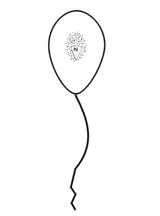
Figure 1: Zoospore of Chytridiales. N=nucleus is surrounded by ribosome (=black dots). The ribosome is loosely associated with the nucleus and is not enclosed within a nuclear cap, which is absent in this order. Zoospore has a single, whiplash flagellum. |
|
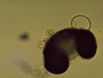
Figure 2: Rhizophydium sphaerotheca: Four visible zoosporangia growing on a pine pollen (one is out of the plane of focus and is behind one of the more visible sporangia). Zoospores can be seen in |
| two of the three visible zoosporangia. The third has released all of its zoospores. Rhizoids (not visible) anchor the zoosporangia to the substrate. | |
This species is known only to reproduce, asexually. Zoospores released from a zoosporangium will swim for a period of time until a suitable substrate, e.g. pine pollen is located. The zoospore attaches itself by producing rhizoids that will grow into the substrate and anchor it. As the cell feeds on the host, growth of the cytoplasm and numerous mitotic divisions will occur, and a cell wall will form around the original zoospore membrane. The cell wall grows as the protoplasm increases in volume. The enlarged cell becomes the zoosporangium and cleaveage of the protoplasm will produce the individual zoospores that are released through a predetermined pore.
Order: Blastocladiales
Fungi in this order are more complex than the
previous order. True mycelium is produced, which is coenocytic. Zoospores in this order
differ from those in the Chytridiales in that the ribosome that is around the nucleus is
enclosed around a nuclear cap, which is an extension of the nuclear membrane (Fig
3).
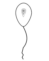 |
Figure 3: Zoospore characteristic of Blastocladiales. Ribosome around nucleus (=N)is enclosed by an extension of the nuclear membrane, the nuclear cap (=NC). |
We will examine Allomyces macrogynus, a well studied species which has been determined to have a true alternation of generations, i.e. a sporic life cycle, with a multicellular haploid and diploid thallus is produced during its life cycle. This type of life cycle is normally associated with plants and with respect to fungi is only known to occur in this order.
The haploid thallus is the gametothallus (=gamete producing thallus), which produces female and male gametangia when mature (Fig. 4). The male and female gametangia occur in pairs and are terminal and subterminal, respectively (Fig. 5). Although not typical, more than a pair of gametangia may occur on each apex of the gametothallus. Male gametes may be distinguished by their orange color, due to carotene pigment, and their smaller size (Fig. 6). Female gametes are colorless and are several times larger than the male (Fig. 6). Thus, this is an example of an anisogamous species. It has long been known that male gametes are attracted to the females by a hormone, sirenin, which is secreted by the female gametes. However, it has recently been demonstrated that a female-attracting hormone is produced by the male (Pommerville, 1990). This hormone has been named parisin.
The diploid thallus is the sporothallus
(=spore producing thallus) which produces two types of zoosporangia (Fig. 7). One is just
called the zoosporangium (=mitosporangium) and the other is the resistant
sporangium (=meiosporangium). Both are usually produced terminally but
are usually not at the same location. However, clusters of the same type of sporangia may
occur. The zoosporangia produces diploid zoospores which germinate to produce more
sporothalli. Thus, the diploid zoospore functions as a means of asexual
reproduction, and as long as the environment remains favorable for the sporothallus, the
zoospores will continue to reproduce in this fashion. Because of the ephemeral habitats in
which the aquatic fungi are usually found, eventually the environment of the sporothallus
becomes unfavorable and the sporothalli will die. However, the resistant sporangia, which
have thick, pigmented walls are resistant to desiccation and will survive after the rest
of the thallus dies. When favorable conditions return, meiosis will occur in the resistant
sporangia and produce haploid zoospores that will germinate to produce the
gametothallus.
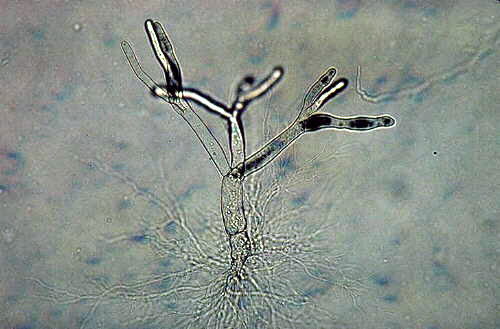 |
Figure 4: A young gametothallus on which the female and male gametangia is produced. The thallus is anchored to its substrate by the extensive rhizoids at the base. |
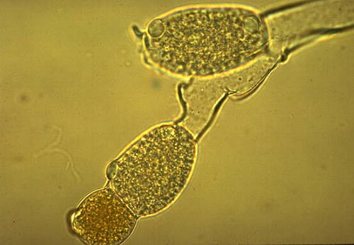 |
Figure 5:Terminal branches of mature gametothallus with one male gametangium two female gametangia visible. Note orange color of male gametangium. |
 |
Figure 6: Male and female gametangia releasing gametes. Note the larger size of female gametes. Thus, gametes are anisogametes. Female and male gametes are attracted to one another by |
| production of the hormones, sirenin and parasin, respectively. | |
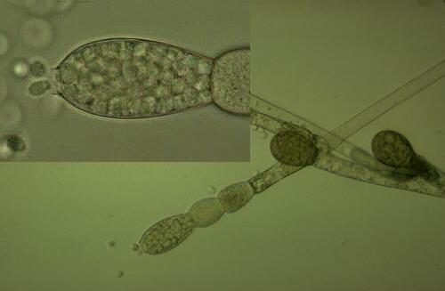 |
Figure 7: Sporothallus with zoosporangia and resistant sporangia. The colorless, more elongated sporangia are the zoosporangia and the dark pigmented, rounder sporangia are the |
| resistant sporangia. Note inset, on upper left, showing zoospore release from zoosporangium. | |
Literature Cited
Bartnicki-Garcia, S. 1970. Cell Wall Composition and Other Biochemical Markers in Fungal Phylogeny. Pp. 81-103. In: Phytochemical Phylogeny. Ed. J.B. Harborne. Academic, London.
Pommerville, J. 1990. Pheromone Interactions and
Ionic Communication in Gametes of Aquatic Fungus Allomyces macrogynus. J.
Chem. Ecol. 16:121-131