Anthocerophyta & Bryophyta
![]() Botany 201 Lab-Hepatophyta
Botany 201 Lab-Hepatophyta ![]()
Anthocerophyta & Bryophyta
The Divisions we will explore today are commonly known as "Bryophytes". One Division is called the Hepatophyta (Liverworts) and includes three Orders that we will examine. These are the Marchantiales, Metzgeriales and the Jungermanniales (Leafy Liverworts). The first two Orders are called Thallose Liverworts because they do not have a leaf/stem organization but consist of flat spreading thalli. I am not so concerned that you recognize the differences between these two orders. The Marchantiales have the most advanced vegetative and reproductive features of the two & you should concentrate your studies on it. The Jungermanniales has leaf/stem organization, hence the designation of "Leafy Liverworts". This should be easy to remember.
The Hornworts (Anthocerophyta) derive their common name from the German name for plant (wort) and their elongated horn-like Sporophyte. They are also thallose in terms of their gametophyte but they have some distinguishing traits that you should recognize.
The "True Mosses" comprise the Division Bryophyta. These have a leaf/stem organizational pattern and are the most familiar members of this triad.
General Features: All of these are land plants (terrestrial) with some aquatic forms. They are very small. The sporophyte and gametophyte have very different morphologies and the sporophyte is usually partly dependent on the gametophyte. Some gametophytes are organized as a thallus. A thallus is a flattened growth form, which does not have discrete organs like stems, leaves and roots. However, some of these have a leaf-shoot organization similar to other land plants.
Gametes are produced in jacketed, multicellular structures (Gametangia). Antheredia produce flagellated sperm, which swim to an Archegonium which, contains an egg. What type of sexual reproduction is this?
The Sporophytes grow out of the archegonia and are usually anchored to the gametophyte by a Foot. Eventually, Meiosis produces haploid spores in the Capsule (Sporangium). Spores germinate and develop into gametophytes. What type of reproductive cycle is this? Gametic, Sporic, Zygotic???
These are relatively simple organisms but they show numerous adaptations, which probably represent some of the key features of the first plants which colonized, land. These include specific modifications of the Epidermis (unicellular external layer) and internal tissues. Some of these organisms have Vascular Tissues & Support Tissues similar to those of other terrestrial plants.
Lab Goals (We will have the same basic goals for all of these labs).
1] Understand the basic anatomy of gametophytes and sporophytes
2] Recognize key differences in the growth patterns and reproductive strategies of gametophytes and sporophytes.
3] Understand how various cell, tissue and "organ" adaptations equip these organisms to survive and reproduce in terrestrial environments.
4] Think about the factors, which limit these organisms in terms of their abundance, growth and reproduction on land.
5] Relate various features which bryophytes share with Algae, especially Chlorophyta
General Lab Procedures
In all of he labs on terrestrial plants we should take an outside to inside approach.
Examine the overall form (morphology) of the organism from stem to stern with your eyes.
Study the organism with a dissecting scope for finer detail.
Examine fresh sections with the light microscope to observe gross anatomical features. (WE will do the sectioning.)
Use stains to identify different chemical aspects of cells & tissues.
Study prepared slides, which illustrate detailed anatomical traits.
Hepatophyta (Liverworts)
The Gametophore (That which bears Gametangia)
The gametophore represents the most conspicuous phase of the bryophyte life cycle. In some cases the gametophore is a specialized structure produced by the gametophyte. In other cases the entire gametophyte is a gametophore. We will study examples of various Taxa to illustrate the variations in complexity achieved by these organisms.
Thallose
liverworts have broad flat vegetative structures called a thallus, and they
do not produce leaf-like protrusions.
Note the Similarities with Chlorophyta (Green Algae)
Locate the Midrib. Hold them up to the light. Are they Translucent? Are they Dorsiventral?
Are Rhizoids present?
Commercial
Slides: Observe the following
Pellia Thallus with Apical Cell.
Pellia Antheridia - Note their position & compare with Marchantia (Next Taxon).
Pellia Archegonia - Note their Position & the presence of Protective Tissue (Involucre)
Pellia Mature Capsule - Note the Elaters.
Also Look carefully at the Spores. Are they Unicellular or Multicellular. If they are multicellular, what type of development do they have?
Why is this a positive reproductive adaptation?

We will examine two species in the Marchantiales (Marchantia & Demortaria).
Observe the overall growth habit of these. They branch dichotomously (in two) at the apex of the thallus. Locate the apex.
Take part of
a thallus and place it on a Petri dish top or bottom & examine it with a
dissecting scope.
Look first at the bottom (ventral) surface to find the rhizoids. Where are they located? How does this affect the appearance (morphology) of the thallus? What do you think they (the Rhizoids) do?
Scrape some off into a drop of water & examine with the light microscope. Are they unicellular or multicellular?
Observe the upper (dorsal) surface with the dissecting scope. Can you pick out any special features? You may need to blot it dry with a Kimwipe.
Do not look
too long! The  answers should be obvious.
Notice that the Thallus is Compartmentalized. Think of the Titanic, then think why this could be advantageous for a plant like Marchantia.
Observe Commercial slides of Marchantia & Fresh Sections prepared by your intrepid and fearless lab instructor.
Commercial
Slides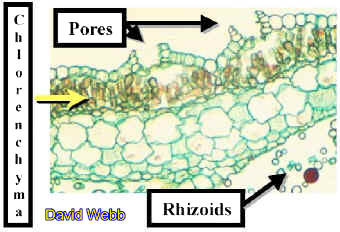
Marchantia
Thallus Serial - Locate the Apex & follow differentiation backwards from it.
Especially note the origin of the scales, rhizoids, pores &
photosynthetic tissues.
Is there an upper & lower Epidermis?
What other Tissues do you see?
How are these organized in the thallus?
Locate the specialized Gametophores that produces either Archegonia or Antheridia. Note its point of origin.
Archegoniophores appear indented in outline while Antheridiophores are less so.
Observe
Commercial Slides of Marchantia Antheridiophores, which produce Antheridia.
Note the jacket layer,  which
surrounds the lattice-like array of spermatogenous cells, which will produce
biflagellate sperm.
which
surrounds the lattice-like array of spermatogenous cells, which will produce
biflagellate sperm.
Examine a commercial slide of Marchantia Archegoniophores, which produce archegonia. Locate the archegonia and identify the neck, neck canal & egg. Be sure to observe the DEMO slide!
Archegoniophore |
Archegoniophore |
Archegonia |
The Sporophyte
Observe commercial slides of Marchantia Sporophytes. Note the various stages of development. What is the ploidy level of the sporophyte????
Identify the Seta, Sporangium, Spores & Elaters.
Examine the sporangium
wall and the elaters.  They
both have wall thickenings, which play a role in spore dispersal.
They
both have wall thickenings, which play a role in spore dispersal.
Observe the DEMO
Slide of Spore Tetrads. What are tetrads? Tetra = four. Why does this number suggest
that an important process has occurred? What is that process?
Locate sporophytes on the archegoniophores of living Marchantia
thalli.
Make a squash of a sporangium, let it dry on a slide.
Place this onto the stage of your microscope and breathe on it while you look through the oculars at 10X.
Jungermanniales (Leafy Liverworts)
Observe living gametophytes.
The liverworts will appear to be flattened compared to the accompanying moss. The leaves have a 2-ranked rather than a spiral appearance.
Note the lateral branching pattern on larger "stems". This is different from the dichotomous branching seen earlier with Marchantia.
Separate several smaller stems and mount them on a large drop of water on a microscope slide.Add a large
cover slip and observe under low power. Locate the apical meristem, and note the
two-ranked appearance of the "leaves". Note the transparency
of the leaves (only 1 cell thick) and the absence of a midrib.
Switch to 10X. Use the condenser iris to see the small, bilobed ventral
leaves. This is an example of anisophylly (unequal leaves). What might be the
functions of these different leaves
Locate Rhizoids on the ventral surface. These will appear like starbursts at intervals along the stem.
Commercial Slides (Demos)Porella Female Branch with Archegonia.
Porella
Sporophyte - Note the Spore walls! Identify two Spore adaptations, which could have
positive Adaptive Value!
Anthocerophyta
(Hornworts) 
Examine the gametophores of Anthoceros and mentally compare them with Marchantia..
Locate the sporophytes,
which develop from sunken archegonia.
Locate various stages of sporophyte development.
The calyptra forms a sheath surrounding young sporophytes. Eventually the sporophyte breaks through the calyptra, elongates and dehisces to release its spores.
DEMO Your intrepid Lab Instructor will make fresh sections of Anthoceros thalli. Note the internal chambers. These are sometimes filled with mucilage and nitrogen-fixing cycnobacteria. Is this an important adaptation???
Also note the chloroplasts. How many are there per cell?? . Note the presence of a round element in each chloroplast. Staining with IKI reveals the presence of starch, which stains blue-black to brown. This is a pyrenoid. Where have you seen this before?? __________________________. What does this suggest to you regarding the origins of these plants?
Commercial Slides
Observe DEMO
of the Sporophyte Foot embedded in the Gametophyte. Note the way in which the cell
walls of the foot have a digitating (finger-like) appearance. What is the functional
significance of this?
Observe DEMO cross-section through the Capsule of the Sporophyte. Note the thick-walled Epidermis, several layers of Photosynthetic Parenchyma & the Spores. These Sporophytes have high photosynthetic rates.
Division Bryophyta
Pogonatum
or Polytrichum 
Note the upright orientation of the stem.
Also note the spiral arrangement of leaves around the stem.
Try to locate male and female gametophytes. We will help with this!
The males
have enlarged, reflexed perigonial leaves just below the apex. Antheredia
are produced within the concave bowl created by the leaves,
which are usually reddish in color. Antheredia can also be red.
concave bowl created by the leaves,
which are usually reddish in color. Antheredia can also be red.
When mature Antheridia are placed in water, many sperm can be observed!!!!!
The stems of female
plants are more elongated and pointed at the apex and surround the
apical meristem like hands folded in prayer. 
The exerted tips of the archegonial necks can be seen protruding from the stem apex of female plants with the aid of a dissecting scope. They resemble little waxy droplets.
Observe the open neck canals through which the sperm must swim to the egg. If you are lucky you may see a young sporophyte emerging from an archegonium.
DEMO We will set up a few male and female plants for you to observe.
Note the massive size of the antheridia and archegonia compared to those of liverworts.
Mosses like Pogonatum which have upright, unbranched stems with terminal gametangia are called Acrocarpous (acro = apex, carpous = reproductive structure)
Pleurocarpous mosses have highly branched, prostrate stems. Leucobryum is an example of a pleurocarpus moss.
Observe a DEMO
various stages of Pogonatum sporophyte development with a dissecting scope.
Remove a mature sporophyte, which has not shed its spores. Locate the seta.
Remove the papery calyptra and find the capsule (sporangium) and operculum.
Use a dissecting needle to pry off the operculum.
Note the
red peristome teeth, which aid in spore dissemination also note, the septum
which blocks spore release.
Mount some spores in water and observe with a compound Microscope. Do they appear to have thick walls? What is their Color? What does this tell you about the resulting Gametophyte plant?
Commercial Slides
Observe transverse sections of Pogonatum stems.
Note the relative complexity of the stem compared to liverworts.
The dark outer layer is the Epidermis. Note the thickness of the cell walls.
The next zone is a broad cortex. Cells near the epidermis have thick walls, while more internal cells have thin walls.Locate the Central Strand.
Thick-walled Hydroids occupy the center of the stem.
Leptoids which have densely-stained contents are present between the hydroids and the cortex.
What do you think are the principal functions of these tissues??????
Observe leaf
cross-sections in the same slide. Note the difference in
anatomy between leaves cut close to the stem and those further away from it.
slide. Note the difference in
anatomy between leaves cut close to the stem and those further away from it.
Note the prominent midrib, which contains several different types of cells in both kinds of section.
Observe the
parallel rows of loosely connected photosynthetic cells.
How does the structural complexity of these leaves compare with Sphagnum and leafy liverworts????
Observe Longitudinal sections of Polytrichum or Pogonotum Capsules. Locate the central Columella & the Sporogenous Cells. Also locate the Annulus & Operculum. What is the function of the Annulus?
Living Material - Observe Mature Capsules, which have released their spores & locate the Peristome. What is its function?
Observe Commercial Slide DEMO of moss Protonema. Note the unicellular nature of the protonema. Can you find Rhizoids?
Observe Protonema with Buds. These are green in nature but may have stained Brown in commercial slides. What do these become?????
![]()