| Anthocerophyta - 2 |
Gametangia are Embedded in the Dorsal Surface.
Biflagellate Sperm are produced by the Antheridia.
The Archegonia are unique and lack a Neck & Venter. (A minor point). |
Sporophytes |
|
 Sporophytes at Various Developmental Stages The Archegonia produce a Calyptra which protects the Sporophyte during early Development. It is translucent at first but later it becomes green. |
|
| Sporophyte DevelopmentThe Sporophytes are Cylindrical & highly elongate. They
grow for a relatively long time and have an Intercalary Meristem like grasses. This means that meristematic activity is confined to the base rather than the apex. They are are not exactly Sequoia-like in growth, but they can reach 16 cm in height. |
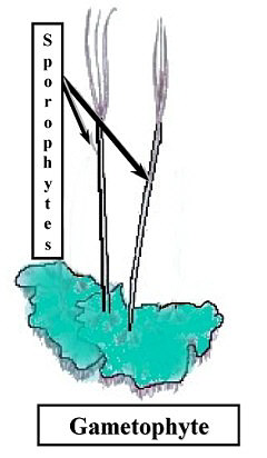 Diagram showing the relationship between Sporophytes & Gametophytes |
Sporogenous Tissue develops in the capsule. The central Columella is sterile and can have cells with spiral secondary wall thickenings. This provides support for the elongated Sporophyte.The Meiospores are surrounded by several layers of Parenchyma cells and an Epidermis. Stomata are present in the Epidermis. |
|
![]()


