of Vascular Plants
![]() Bot 201 Lab-Vegetative Features
Bot 201 Lab-Vegetative Features
![]()
of Vascular Plants
The purpose of this lab is to acquaint you with the various types of tissues found in Vascular Plants and to show you how they are organized to form a "Typical Vascular Plant". This may seem daunting at first until you realize that basic plant anatomy is very simple.
Cell
Types & Tissues
Meristematic Tissue
Meristematic
tissue produce all the cells in herbaceous plants. The cells originate in the Root or
shoot Apical Meristems. Meristematic Cells are Spherical
(Isodiametric) and densely cytoplasmic. They have a relatively large Nucleus,
few Vacuoles and thin Cell Walls. Consequently, they
readily absorb biological stains and appear as dense areas in the apices
of Roots and Shoots. The Apical Meristem in Roots is actually subterminal because it is
covered by a Root Cap. The root cap protects the delicate meristem cells and it also
secretes carbohydrates which lubricate the root
They have a relatively large Nucleus,
few Vacuoles and thin Cell Walls. Consequently, they
readily absorb biological stains and appear as dense areas in the apices
of Roots and Shoots. The Apical Meristem in Roots is actually subterminal because it is
covered by a Root Cap. The root cap protects the delicate meristem cells and it also
secretes carbohydrates which lubricate the root  as
it grows through the soil.
as
it grows through the soil.
There are two basic types of Apical Meristems. Nonseed plants have a large Apical Cell which gives rise to the plant body. Seed plants have Multicellular Apical Meristems which function as a unit.
Primary Tissues
There are Three Basic Tissues that comprise all herbaceous (soft bodied) plants. These are Dermal, Vascular & Ground.
Plants are
made like reinforced concrete. There is a outer mold,
steel rods and concrete which fills in the rest of the
volume.
There is a outer mold,
steel rods and concrete which fills in the rest of the
volume.
The first tissue is the Epidermis which is typically on the surface (Dermal) and is usually one cell thick. If it is on the surface it is probably Epidermis. This is like the mold for the concrete.
The
next is Vascular Tissue. It is typically found in longitudinal
columns. There may be one large central  column, a ring
of smaller columns or multiple rings of small columns. These columns are
the steel rods.
column, a ring
of smaller columns or multiple rings of small columns. These columns are
the steel rods.
Finally, there is the Ground Tissue which occupies the rest of the plant organ. This is like the concrete above. I regard the Ground Tissue as BACKGround Tissue.
Cross Section of a Coleus Stem: Note the large Vascular Bundles in each corner of the stem (steel rods). Everything else is Ground Tissue (Concrete) except for the Epidermis (Mold) which forms the surface layer of cells. |
 |
The Epidermis is readily identified by its location.
Vascular Tissues, especially Xylem, have strong characteristic traits which make them relatively easy to identify.
If it is neither Epidermis nor Vascular Tissue, it must be Ground Tissue.
There are two
Vascular Tissues, Xylem & Phloem.
The conducting cells in the Xylem are called Tracheary
Elements. These have thick walls that stain red
due to the presence of Lignin. Lignin makes their cell walls watertight,
inflexible and strong. Consequently, Tracheary
Elements have two main
functions, water transport and structural support.
They usually have a hollow appearance because they are dead at maturity.
These traits make Xylem relatively easy to spot in a cross section.
main
functions, water transport and structural support.
They usually have a hollow appearance because they are dead at maturity.
These traits make Xylem relatively easy to spot in a cross section.
Phloem
is associated with Xylem. Phloem cells do not generally have diagnostic
traits like xylem but they can usually be discerned by their position. They have
relatively thin cell walls and have a narrower diameter than the largest
Tracheary Elements. In some cases, Phloem exhibits a definite
pattern of small cells and large cells. The large cells are Sieve
Elements (S). 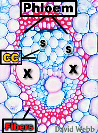 These are the cells that are specialized for Sucrose Transport.
The smaller cells are Companion Cells (CC). These
regulate the physiological activities of the Sieve Elements. Phloem contains
Callose. Callose regulates the size of the openings between adjacent Sieve
Elements. Callose is a carbohydrate that stains with Aniline Blue and is fluorescent under
Violet light. This is the best way to verify the presence of Phloem because Callose is not
present in other cell types.
These are the cells that are specialized for Sucrose Transport.
The smaller cells are Companion Cells (CC). These
regulate the physiological activities of the Sieve Elements. Phloem contains
Callose. Callose regulates the size of the openings between adjacent Sieve
Elements. Callose is a carbohydrate that stains with Aniline Blue and is fluorescent under
Violet light. This is the best way to verify the presence of Phloem because Callose is not
present in other cell types.
 Isodiametric Parenchyma Cell containing Chromoplasts: Each red dot is a Chromoplast that Contains Carotenoids. |
 Elongate Palisade Parenchyma with Chloroplasts |
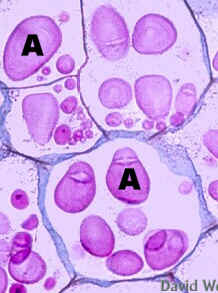 Parenchyma from Potato with large Amyloplasts: This is a commercial slide. |
 Parenchyma Cells containing Amyloplasts. This was stained with IKI which has stained the Starch Brown. |
There are two major types of Ground Tissue (Parenchyma & Sclerenchyma).
Parenchyma cells can be Isodiametric (equal size in all directions) to elongate in shape. They have thin cell walls which contain a lot of hydrophilic pectins which have a characteristic staining reaction. They usually have well developed Plastids like Chloroplasts or Amyloplasts. Amyloplasts store starch.
Sclerenchyma
cells are usually elongate in shape 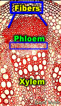 and have thick walls impregnated with Lignin.
They stain red in most prepared slides due to their Lignin
content. Lignin makes cell walls extremely strong
and inflexible. This makes Sclerenchyma a good support
tissue. Sclerenchyma is usually associated with Vascular
Tissues and may completely surround them.
and have thick walls impregnated with Lignin.
They stain red in most prepared slides due to their Lignin
content. Lignin makes cell walls extremely strong
and inflexible. This makes Sclerenchyma a good support
tissue. Sclerenchyma is usually associated with Vascular
Tissues and may completely surround them.
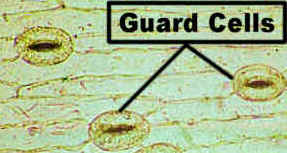
There are a wide variety of Epidermal cell types. The typical Epidermal Cell resembles Parenchyma but it has only tiny plastids. All Epidermal Cells have a waxy Cuticle on their outer surface. They may contain Red/Purple Anthocyanins in their vacuoles. This gives the cells a uniform color. Chloroplasts are present in the Guard Cells which are part of the Stomata. The Guard Cells tend to be smaller than typical Epidermal Cells, and they occur in discernable patterns. The Epidermis can produce a wide range of hairs (Trichomes). These can be unicellular or multicellular. They may absorb water from the soil, secrete chemicals to the surface, inject chemicals into the skin, trap & digest insects and perform many other functions.
 Surface View of Epidermis from a Leaf: Note the undulating Epidermal Cells plus the Stomata (S) and Trichomes (T). |
 Lily Epidermis showing the Elongate Epidermal cells and the Guard Cells which are part of the Stomata |
 Surface of Kukui Leaf with branched Trichomes |
 Isolated Kukui Trichome |
 Cross Section of Venus Fly Trap Leaf: Note the Epidermal Layer and the Multicellular Trichomes which are part of the Epidermis. |
 Epidermis of Silver Sword showing the Guard Cells (GC) of the Stomata |
Trichomes are most abundant with flowering plants! Other Divisions generally lack hairs, except for root hairs.
Tissue Organization and Organs
The most
simple pattern of tissue organization is  seen in Roots. Imagine a
stack of ice cream cones. Fill the first cone with raspberry ice cream.
This is the Xylem. Coat the inside of a second cone with a layer of blueberry
ice cream. The ice cream represents the Phloem. Place this over
the first cone. Coat another cone with mint ice cream and place it over
the other two cones. The mint ice cream is ground tissue.
We should have a stack of three cones. The outermost cone shell is the Epidermis.
seen in Roots. Imagine a
stack of ice cream cones. Fill the first cone with raspberry ice cream.
This is the Xylem. Coat the inside of a second cone with a layer of blueberry
ice cream. The ice cream represents the Phloem. Place this over
the first cone. Coat another cone with mint ice cream and place it over
the other two cones. The mint ice cream is ground tissue.
We should have a stack of three cones. The outermost cone shell is the Epidermis.
Some Stems and Leaves have the same kind of organizational pattern!
 The Root and some Stems have concentric circles of Epidermal Ground & Vascular Tissues |
 Some Leaves have only one Vascular Bundle. These are called Microphylls. |
 Leaves of Monocots like Sugarcane have many Vascular Bundles of similar dimensions. |
 Dicots like Ohi'a Lehua have a large central Vascular Bundle called a Midrib, plus smaller and smaller Lateral Bundles. |
 Monocot Stems have many Vascular Bundles arranged in a complex pattern within the Ground Tissues. |
 Dicot Stems have one ring of Vascular Bundles located in the periphery of the Ground Tissues. |
Lab Activities
Meristematic Tissue
Examine Longitudinal sections from Root and Shoot tips. Locate the Apical Meristems and note their general features with your 40 X Objective.
Follow cell files as you scan towards the base of each Apical Meristem & look for signs of Cell Enlargement and Differentiation (changes in the Cell Walls).
Epidermis
Observe Commercial Slides of Lily Epidermal. Note the Guard Cells of the Stomata. The Guard Cells can open and close to regulate gas exchange with the atmosphere. Also note the elongate Epidermal Cells
 |
 |
| Lily Epidermis | Epidermis with an extremely thick Cuticle! |
Observe a Demonstration slide which shows an Epidermis with an extremely thick Cuticle.
Take
a Pigmented Coleus Leaf.
Cut a 2 x 2 cm square piece from the middle of the leaf.
Place this upside-down on a Microscope Slide.
Place this on the stage of your microscope and move it into the light path.
Use the 4 X Objective but flip up the High Power Condenser Lens. This should produce a small spot of light
Turn the illumination to MAXIMUM!
Do NOT Look through the Objectives when you do this. The intense beam could damage your eyes if it is not blocked by your specimen.
Move the thin part of the leaf into the light path.
Focus up and down until you see details of the Epidermal surface.
You may switch to the 10X objective to see more details.
You should be able to see Trichomes & Pigmented Epidermal Cells & Guard Cells. The latter may be a little hard to find.
Do NOT spend too much time on this. The goal is to give you an appreciation of Epidermal complexity
Dicot
Root
Observe Intact Water Hyacinth Roots. The hair-like projections are actually Lateral Roots. Also note the prominent Root Cap. This protects the delicate Root Apical Meristem. The latter is the source for the Root Cap and the Root Body (everything except the cap)
Observe Commercial Slides of Root Cross Sections.
Locate the Epidermis, Ground Tissue and Vascular Tissues.
The Ground
Tissue is composed of Parenchyma cells. These have Thin
Cell Walls and a round shape. They may have numerous, 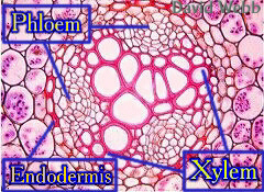 starch-storing Amyloplasts.
Carefully examine the Vascular Tissues and Identify Phloem & Xylem.
The Xylem Stains Red due to the presence of Lignin and has a star-like shape.
The Phloem lies between the radiating arms of the xylem. Its cells are much smaller than those in the xylem and they have very thin walls.
The Endodermis is the innermost part of the Ground Tissue and forms a boundary layer between the Vascular Tissues (Stele) and the Ground Tissue. The Endodermis regulates the movements of water and solute between the Stele and the rest of the root. Some Endodermal Cells have Thick, Lignified Walls while others have relatively thin walls. The Endodermis is one of the most important adaptations of land plants. We will explore this topic later.
Dicot Stem
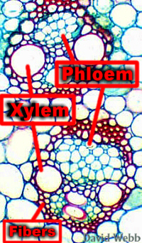
Observe a Commercial Slide of a Typical Dicot Stem and locate the Epidermis, Vascular Bundles & Ground Tissues.
 Typical Dicot Stem: Note the Ring of Vascular Bundles. The Fibers and Xylem stain Purple-Red due to the presence of Lignin |
 Typical Dicot Vascular Bundle |
The Ground Tissue in the center of the stem is called Pith. It contains Parenchyma Cells.
Examine the Vascular Bundles at higher magnification and locate the Xylem, Fibers and Phloem. Phloem lies between the fibers and the xylem and may not have prominent features.
The Ground Tissue between the Fibers and the Epidermis is called the Cortex.
Examine Commercial Slides of Cucumber Stem and locate the Vascular Bundles.
Locate the Phloem which occurs on both sides of the Xylem in Cucumber. Examine the Phloem carefully and try to locate the Sieve Elements & Companion Cells.
The Companion Cells are small and stain darkly while the
Sieve Elements have a larger diameter and have pink
contents.
 Cross Section of Cucumber Stem |
 Vascular Bundle from Cucumber: Note the presence of Phloem on both sides of the Xylem |
 Cucumber Phloem. The small dark cells are the Companion Cells. The larger cells with Pink Contents are the Sieve Elements. |
|
 Highly magnified image of Cucumber Phloem |
 Sieve Plate from a Commercial Slide: You may have trouble finding one like this. We will have a Demo for you to see. |
Try to locate Sieve Plates. These occur on the endwalls of the Sieve Elements and have relatively large openings called Sieve Pores. Sieve Pores facilitate the movement of sugar solution through the Sieve Elements.
Monocot Stem
Examine Commercial Cross Sections of Corn, Sugarcane or Bamboo.
The Vascular Bundles are easy to identify because of the Large Xylem Tracheary Elements.
 Cross Section of Asparagus Stem: Note the distribution of Vascular Bundles throughout the surrounding Ground Tissue
|
 Vascular Bundles from Corn |
Note
the Distribution of Vascular Bundles throughout the Ground Tissue. There
is no Pith in most monocots.
Examine a large Vascular Bundle at higher magnification and locate the Xylem, Phloem and Fibers which surround the vascular tissues.
The Phloem has a geometric organization which makes it easy to identify the Companion Cells and Sieve Elements.
Monocot Leaves
Given the number of Vascular Bundles in monocot stems, it should not be a surprise to see the numerous veins in their leaves.
Examine the leaves of a typical monocot like Sugarcane or ti & note the many veins which run the length of the leaves. Each vein contains one Vascular Bundle. You may see that there are large and small veins which alternate in a regular pattern.
 Cross Section of a Corn (Zea mays) Leaf: Note the Numerous Vascular Bundles. |
Examine Commercial Cross Sections of Sugarcane Leaf and note the following, Upper and Lower Epidermis, Vascular Bundles and Ground Tissue.
The Ground Tissue in a leaf can be called Mesophyll (Middle Leaf). Cells which contain Chloroplasts are called Chlorenchyma or Photosynthetic Parenchyma.
Cross Section of Sugarcane Leaf: |
Dicot Leaves
Dicot Leaves usually have a large central Midrib which contains a large Midvein (Vascular Bundle). Minor Veins branch from the midvein at oblique angles. Consequently, the minor veins may be asymmetrical in cross sections.
Examine a typical Dicot leaf like Kukui and note the Vein Pattern (Venation). You may need to scrape of some hairs to see the lower surface. Mount the hairs in water and examine them under the microscope. Layteral veins get progressively smaller and smaller. This is called Reticulate or Net Venation.
Monocots have "Parallel" Venation because the Vascular Bundles appear parallel to one another and look unbranched. However, close scrutiny shows that they produce Lateral Veins at angles approaching 90 degrees. Thus, they have a form of reticulate venation called Striate. The Important Point to remember is that Monocots and Dicots have Reticulate Venation.
They are both MEGAPHYLLS (big leaf) because they have more than one Vascular Bundle.
Examine a Commercial Cross Section of a typical Dicot Leaf like Pear (Pyrus).
Examine the Midrib and locate the Xylem & Phloem.
 Cross Section of a Typical Dicot Leaf: Note the Vascular Bundle or Midvein). Also note the large air spaces in the Ground Tissue (G) which is part of the Blade (Lamina) |
 Cross Section through the Blade or Lamina of a Dicot Leaf: Note the Variety of Cell Types which have specialized functions. |
Dicot Leaves Typically have two kinds of Mesophyll (Palisade & Spongy). The Palisade cells are like columns and are tightly packed. Spongy cells may be highly branched and are loosely packed. The Palisade Cells intercept most of the light and perform most of the Photosynthesis. The Spongy Cells also contain chloroplasts but mainly serve as a gas reservoir. Note the Lateral Vein.
![]()

