Vegetative Growth
Leaves: Angiosperms produce a wide array of leaves. These come in a vast array of sizes and shapes. The only other Division that approaches the Anthophyta in terms of variation in Leaf morphology & structural complexity is the Pterophyta. Angiosperm leaves are Megaphylls. This means that they have more than vein in each leaf. Palms produce the largest leaves. They may also be the most complex in terms of their venation.
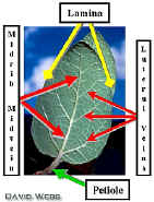
Dicot Leaves are connected to the stem by Petioles. Many Monocots do not have petioles but have a distinct Sheath which connects the Blade to the Stem.
 |
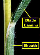 |
| Typical Dicot Leaf with Reticulate Venation | Base of a Monocot Leaf: Note the Sheath which connects the Blade to the Stem |
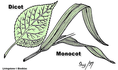
There are two basic patterns of Venation. Dicots have Net (Reticulate) Venation. This is characterized by a network of veins which branch such that each branch becomes smaller and smaller. The smallest veins circumscribe small regions of the blade called Areoles.
Monocots have "Parallel" or Striate venation which is characterized by veins which run parallel to one another over a large expanse of the Blade (Lamina). Close inspection shows that the Parallel Veins are interconnected by small lateral veins. Consequently, monocots also have a network of veins.
Observe samples of intact Dicot and Monocot Leaves and try to decipher their Venation.
Observe Demos of cleared monocot and Dicot Leaves and compare these with intact specimens.
Leaf Anatomy: We looked at leaf anatomy in the first lab on Land Plants. Leaves consist of an upper and lower Epidermis, Vascular Bundles and Ground Tissue called Mesophyll. Due to the major differences that can occur between monocots and dicots cross sections can have characteristic appearances. Dicot leaves tend to have a thick midrib and a thin Lamina. Monocots usually do not have a midrib and the blade is more uniform in its thickness. Because the large veins in monocot leaves lie parallel to one another, they are cut at a 90 degree angle in a cross section. Consequently, they produce a highly organized profile. Because branch veins in dicot leaves depart at oblique angles, they are usually cut at an oblique angle in cross sections. Most Dicots have a Palisade arrangement of Mesophyll tissues. While monocots either have a uniform Mesophyll or one in which the Photosynthetic Parenchyma are distributed like spokes on a wheel around the Vascular Bundles. This is called Kranz (wreath) Anatomy and signifies that these plants have C4 Photosynthesis. Sugarcane is an example of the latter. Some dicots also have C4 Photosynthesis.
We will give you a Typical Dicot and a Typical Monocot leaf to examine. Cut two (2 x 2 cm) squares from each and examine them with your compound microscope to see surface features, especially stomata. Do the stomata occur on both sides of the leaf or only on one side? Why might this be important?
Refresh your memory by examining cross sections of typical Dicot and Monocot leaves. Identify the three major tissues and try to determine whether the leaf has Kranz Anatomy.
Stems: The basic difference between the stems of Monocots and Dicots is the distribution of Vascular Bundles. Dicots have one ring of Vascular Bundles in the outer region of the stem. Consequently, there is a distinct Pith. The Vascular Bundles of Monocots are distributed throughout the Ground Tissue such that there is no Pith.
Observe Cross Sections of Typical Monocot and Dicot Stems.
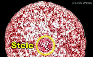
Roots: The Primary Root has not exhibited a lot of variation over the course of Evolution. However, Monocot roots can often be distinguished from Dicot roots. Dicot Roots have a central core of Primary Xylem that is star-shaped. The star has from 3 - 5 rays or arms, and there is NO PITH. Monocot Roots have 10 or more Xylem Rays and there is a Pith in many cases. Phloem alternates with the xylem arms in both cases.
| Cross Section of a Typical Dicot Root | Stele from a Typical Dicot Root |
 |
 |
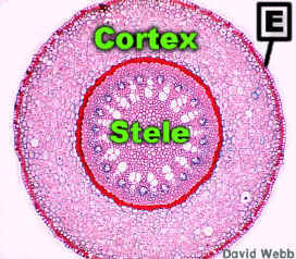 |
 |
| Smilax is a Typical Monocot Root | Vascular Tissues in a Smilax Root |
Observe Commercial Slides of a typical Monocot and Dicot Root
Secondary Growth is one of the most important plant adaptations. Gymnosperms exhibited extensive Secondary Growth. This allowed them to attain great heights and compete for sunlight vertically. Secondary growth also provided protection for plant organs so that they could persist over a long time span.
The Basic Steps in the formation of Secondary Vascular Tissues are illustrated below.
We will use Coleus as a Model plant to study the early stages of Secondary Growth in Angiosperms.
Examine Commercial Slides of Coleus stem and locate the Large Vascular Bundles in the Corners of the stem.
Locate the Interfascicular Region between the Vascular Bundles and look for evidence of meristematic activity.
Locate the Cortex and Epidermis.
Examine the subepidermal cells in the Cortex for Meristematic Activity.
 |
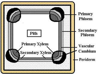 |
| Diagram of a Coleus Stem at the end of Primary Growth | Diagram of a Coleus Stem after a short period of Secondary Growth |
Examine Fresh sections taken from the top of a Coleus Stem. Stain this with Phloroglucinol which will stain the Xylem and Fibers Red due to the presence of Lignin. The Phloem does not stain but it is located between the Fibers and the Xylem.
Locate the Large Vascular Bundles which occur at the corners of the Stem. Is there any evidence of Secondary Growth in the regions between the large bundles?
Use Polarizing Filters to help visualize the Tissues.
Also locate the Epidermis and examine the subepidermal region. Is there any evidence of Periderm formation?
Examine a section from the base of the stem stained with Phloroglucinol. You should see evidence of Secondary Growth by the Vascular Tissues. There should be an unbroken ring of Secondary Xylem that separates the Cortex from the Pith. The ring will be thicker in the corners because this is where the Primary Xylem was located before Secondary Growth occurred.
There is little Secondary Phloem.
Periderm development should be evident in the subepidermal region of the Cortex.
Trees produce large amounts of Secondary Xylem & Phloem. We will use Tilia americana to identify Secondary Tissues.
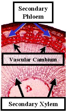 Cross Section of a one year-old Tilia Stem: Note the extensive Secondary Phloem & Xylem. |
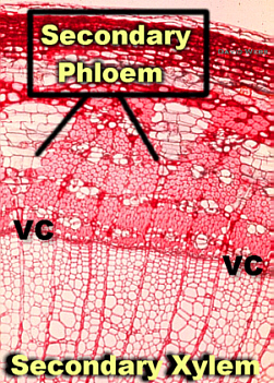 Secondary Xylem & Phloem of Tilia: VC = Vascular Cambium |
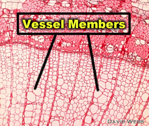 Tilia Secondary Xylem: Note the relative complexity in the secondary xylem of Tilia compared to Gymnosperm Secondary Xylem. |
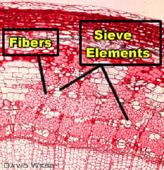 Tilia Secondary Phloem: Note the many Fibers which protect the delicate Sieve Elements. |
| Examine a Commercial Slide of Periderm. Locate the Cork Cambium (Phelogen) and Cork Cells (Phellem). | 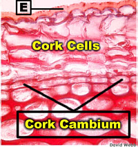 Typical Early Periderm: E = Epidermis |
Examine a Large Specimen from a tree that shows Secondary Xylem & Phloem plus Periderm.
Observe Demo Slide that shows a Root at the end of Primary growth and compare it with a woody Root that has produced a lot of Secondary Growth. Secondary Xylem will comprise most of the root volume.
Some Monocots have Secondary Growth but this is beyond the scope of this course.