Angiosperm means covered seed. The flower is the most important adaptation for the sexual reproduction of plants. The hallmark of angiosperm reproduction is the Carpel.
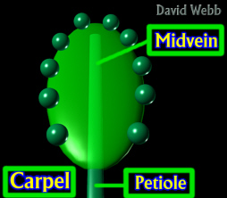
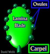 Model of a Primitive Carpel: This is a leaf that bears Carpels along its Margin |
|
|
|
|
|
The
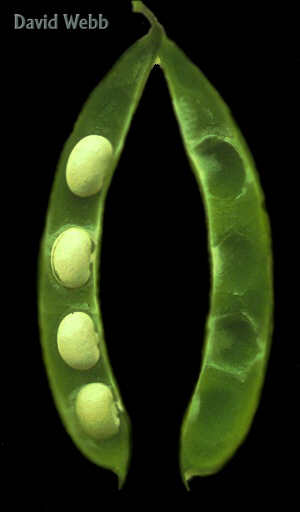
carpel is a highly modified leaf which bears Ovules. To grasp
this, imagine a pea pod. The peas are inside. Carefully slice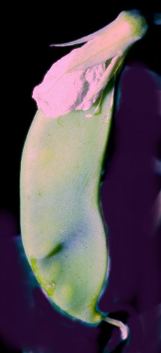 along one margin with a knife, and unfold the fruit.
Voila!!!! What do you see??? A leaf!!!! A Carpel!!! Now, imagine what would happen if you
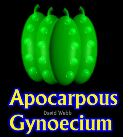
fused several of these together!!! The carpels constitute the Gynoecium.
along one margin with a knife, and unfold the fruit.
Voila!!!! What do you see??? A leaf!!!! A Carpel!!! Now, imagine what would happen if you
fused several of these together!!! The carpels constitute the Gynoecium.
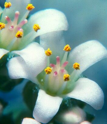
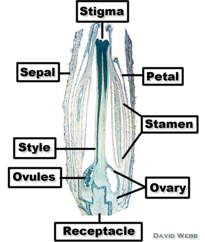
The flower also has an Androecium (Stamens), a Corolla (Petals) and a Calyx (Sepals). These also play roles in sexual reproduction.
One of the goals of this lab is to understand the basic organs which are found in flowers.
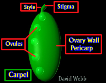
We saw
that the Ovules of Gymnosperms were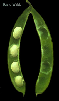 exposed to the atmosphere even
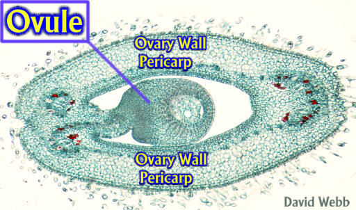
if they were tucked away in a crevice. The angiosperm ovule is housed within the ovary
wall (Pericarp). This provides an extra measure of protection
for ovule and seed development. It may also provide adaptations which help in seed
dispersal.
exposed to the atmosphere even
if they were tucked away in a crevice. The angiosperm ovule is housed within the ovary
wall (Pericarp). This provides an extra measure of protection
for ovule and seed development. It may also provide adaptations which help in seed
dispersal.
One goal for the lab is to link flower structure to fruit type.
We saw the tremendous reduction in complexity with the Gametophytes of Gymnosperms. Further simplification occurs with Angiosperms.
One of the major goals of this lab is to understand the basic outcomes for the gametogenesis of angiosperms.
We will have several simple flowers for you to dissect.
Make sure you find all of the reproductive structures as well as the sepals and petals.
Note any instances of Coalescence or Adnation.
 |
 |
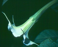
| Datura | Hibiscus |
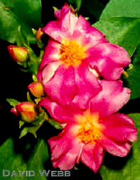 |
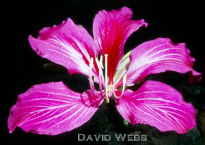 |
| Pereskia | Bauhinia |
 |
 |
| Erythrina | Rubus |
Determine whether the Gynoecium is composed of many individual Carpels, one Carpel or several fused Carpels
Is the Ovary Superior or Inferior?
Make a cross section through the Ovary to locate the Ovules.
Identify the Pericarp (Ovary Wall)
Androecium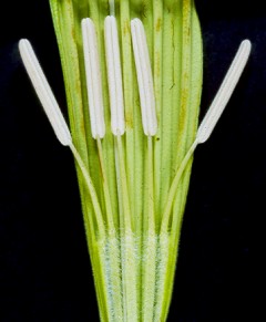
Locate the Stamens
Tease apart the Anthers
Make a wet mount of Pollen and Examine with the light microscope.
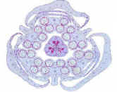
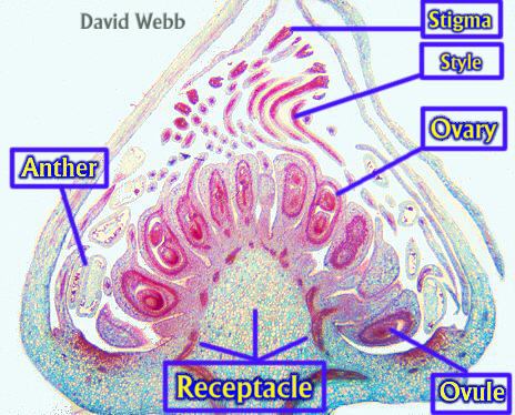
Examine cross sections of Lilium Flower Buds & Locate the following
Sepals
Petals
Anthers
Carpels
Ovules
How many carpels do you observe?
We only have a few full sets of these. We will need to work togeter. I will have three slide boxes which each have one set of slides for Megasporogenesis or for Microsporogenesis. Each Table should take one box & study one of the two processes as a group. Return this slide box, and take a box for the other process.
The scheme that I gave you for the lecture is a lie!!!! Megasporogenesis in Lilium is rather complex and is a bad choice in terms of learning about this. I actually used the Polygonum type. I don't want to go into the Lilium type, so we will put out the slides you need to examine in order to understand what is called Monosporic Development. This refers to the fact that only one Megaspore survives and it produces the Megagametophyte (Embryo Sac).
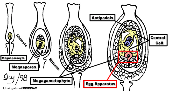
Ovules are produced in the Ovary
of the Carpel. 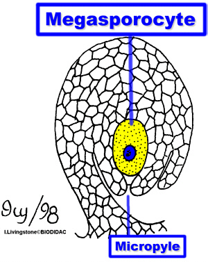 One cell, near the
Micropyle, becomes enlarged and ultimately produces four Megaspores. The enlarged cell is
called the Megasporocyte. Cell that produces Megaspores. The tissue from which it came is
called the Nucellus.The Megasporocyte produces four haploid Megaspores. Three of these
die. The "functional" Megaspore enters Mitosis and produces Eight Haploid
Nuclei. This is followed by Cell Formation. This multicellular structure is the
Megagametophyte or Embryo Sac. Three cells develop near the Micropyle. This is the Egg
Apparatus and consists of the Egg and two Synergids. Three similar cells form at the
opposite pole of the Megagametophyte and are called the Antipodals. The remaining two
nuclei form a large Central Cell. Following Double Fertilization, the Egg forms the Zygote
and the Central Cell becomes the Endosperm. The Endosperm is consequently 3N in this case.
One cell, near the
Micropyle, becomes enlarged and ultimately produces four Megaspores. The enlarged cell is
called the Megasporocyte. Cell that produces Megaspores. The tissue from which it came is
called the Nucellus.The Megasporocyte produces four haploid Megaspores. Three of these
die. The "functional" Megaspore enters Mitosis and produces Eight Haploid
Nuclei. This is followed by Cell Formation. This multicellular structure is the
Megagametophyte or Embryo Sac. Three cells develop near the Micropyle. This is the Egg
Apparatus and consists of the Egg and two Synergids. Three similar cells form at the
opposite pole of the Megagametophyte and are called the Antipodals. The remaining two
nuclei form a large Central Cell. Following Double Fertilization, the Egg forms the Zygote
and the Central Cell becomes the Endosperm. The Endosperm is consequently 3N in this case.
We will set out slides for you to review this as well.
Sporogenous cells occur in the
Pollen Sacs of the Anther. These are called Microsporocytes. These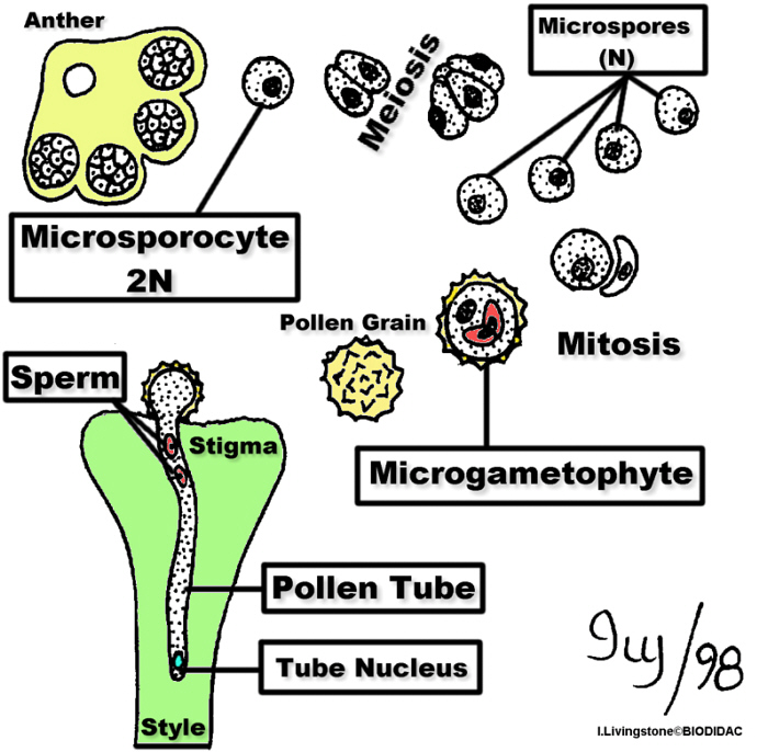 undergo meiosis and produce four haploid Microspores.
Each Microspore undergoes Mitosis such that there are two nuclei in one cell. One of the
nuclei divides again to produce the nuclei of each Sperm. When cell formation is complete
there is one large cell (Tube Cell) and two smaller cells called Sperm. The Sperm cells
float in the cytoplasm of the Tube Cell. The Microgametophyte is known as a Pollen Grain!
When Pollen lands on the Stigma of a receptive carpel it germinates. The Tube Cell
produces the Pollen Tube and the Sperm are carried forward as the tube elongates. The Tube
Nucleus is usually near the tip of the Pollen Tube and the Sperm are in a more basal
position. The Pollen Tube may cover a relatively enormous distance to reach the Egg. The
silk strands on corn cobs are the Stigmas and Styles of individual Carpels.
undergo meiosis and produce four haploid Microspores.
Each Microspore undergoes Mitosis such that there are two nuclei in one cell. One of the
nuclei divides again to produce the nuclei of each Sperm. When cell formation is complete
there is one large cell (Tube Cell) and two smaller cells called Sperm. The Sperm cells
float in the cytoplasm of the Tube Cell. The Microgametophyte is known as a Pollen Grain!
When Pollen lands on the Stigma of a receptive carpel it germinates. The Tube Cell
produces the Pollen Tube and the Sperm are carried forward as the tube elongates. The Tube
Nucleus is usually near the tip of the Pollen Tube and the Sperm are in a more basal
position. The Pollen Tube may cover a relatively enormous distance to reach the Egg. The
silk strands on corn cobs are the Stigmas and Styles of individual Carpels.
Microscopic Flower Buds
Observe Commercial Slides of Various Flower Buds and Locate the Floral Parts
| Tomato
Flower Bud Syncarpous |
Rubus
Flower Bud Apocarpous |
 |
 |
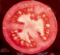 Tomato Fruit: The solitary Ovary has produced one Fruit. This is a Simple Fruit |
 Rubus Fruit: The Ovary of Each Carpel has formed a Fruitlet. This is an Aggregate Fruit. |
Observe
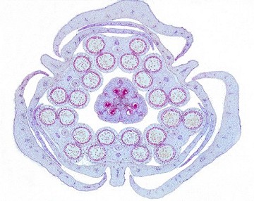
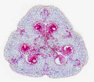
Cross-Sections of Bean (Phaseolus) Flower Buds & Locate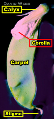
Calyx (outermost) structure
Corolla
Fused Filaments of the Stamens + One free Filament
Ovary Wall & Ovule
Observe DEMO Longitudinal section of a Bean Carpel & Locate
Receptacle
Pericarp
Ovule
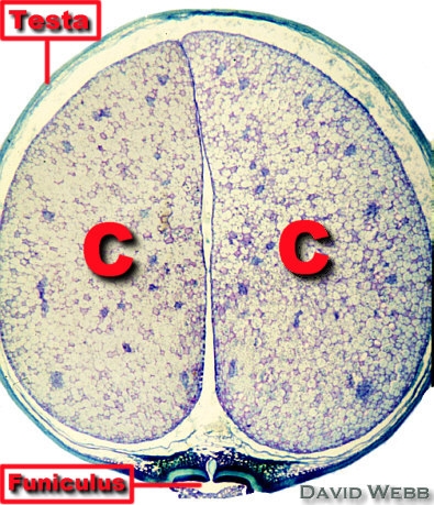
Observe DEMO of a Mature Bean Fruit with Seed & Locate
Ovary Wall
Seed Coat (Testa)
Cotyledons (C)
The Embryo & Seed
The mature ovule is a seed. We do not have enough time to look at seed structure in depth but you should observe Demo Slides showing Lilium & Capsella seeds with Embryos.
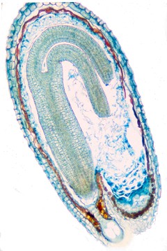 Capsella Embryo (Dicot) |
 Lilium Embryo (Monocot) |
Identify the Seed Coat,
Endosperm and Embryo for both.
Identify the Cotyledons, Shoot Apical Meristem Hypocotyl and Root Apical Meristem for
Capsella.
Fresh Fruit
The Mature Ovary is the Fruit. We will have a small number of fruits for you to examine. Try to locate any residual floral structures that might help you determine whether the fruit is Simple, Aggregate Multiple, Accessory.
Additional Demos
Lilium Young Flower Bud (Longitudinal Section)
Lilium Early Ovule (Find the Megasporocyte a.k.a. Megaspore Mother Cell)
Lilium Pollen (Whole Mount)
Lililu Pollen with Pollen Tubes
Lilium Stigma with Pollen Tubes (The Style is hollow and has a secretory Epidermis)