|
|
Quiz Me! |
Cell structure and function |
|
|
|
Lecture Index | ||
|
|
Course Index |
Last revised: Tuesday, October 19, 1999
Reading: Ch. 7 in textNote: These notes are provided as a guide to topics the instructor hopes to cover during lecture. Actual coverage will always differ somewhat from what is printed here. These notes are not a substitute for the actual lecture!Copyright 1999. Thomas M. Terry
Microscopy
Units of Measurement
- centi- (10 -2 )
- milli- (10 -3 )
- micro- (10 -6 )
- nano- (10 -9 )
- Ångstrom (10 -8 cm., 0.1 nm)
- I'd like to review dimensions before I proceed
Size and Scale
- View " How Big? " from Cells Alive
- View Hyperspace tour at the Biology Place.
Resolving Power
- measures the ability to distinguish small objects close together
0.5 (lambda) r.p. = ______________ (N sinØ)where lambda = wavelength of illuminating light.
- for light scope, can improve R.P. by making lambda smaller or sinØ larger.

- R.P. is smallest for violet light, but because human eye is more sensitive to blue, optimal R.P. is achieved with blue light (~450 nm). Use filters to remove other light in best microscopes
- n sinØ is called numerical aperture . It measures how much light cone spreads out between condenser & specimen. More spread =better resolution. Ø = angle of light cone; maximum value is 1.0
- n = refractive index. n = 1.0 in air. Can --> with certain oils (up to 1.4),called immersion oil.
note: N.A. is property of lens. Look on side of lens to see- Theoretical limit of R.P. for light scope is 0.2 �m.
Optical Instrument Resolving Power RP in Angstroms Human eye 0.2 millimeters (mm) 2,000,000 A Light microscope 0.20 micrometers (�m) 2000 A Scanning electron microscope (SEM) 5-10 nanometers (nm) 50-100 A Transmission electron microscope (TEM) 0.5 nanometers (nm) 5 A Light Microscopes
- Bright-field Light Microscope
- Theoretical limit of R.P. for light scope is 0.2 um.
- Most readily available microscope, found in every biology lab, hospital
- Most biological materials lack contrast; need to fixed and stained
- Thick materials must also be embedded in paraffin or plastic and sectioned
- Phase-Contrast Light Microscope
- Phase scope converts slight diffs. in refractive index and cell density into variations.
- Advantage: can see live material w/o staining
- Fluorescence Microscope
- Fluors are chemicals that adsorb light to produce excited electrons, later reradiate light = flourescence.
- To use, need special type of microscope. Illuminate with ultraviolet or violet light (--> excited fluor). Need filters to remove this light from light traveling to ocular lens; only fluoresced light emitted from object will then appear to eye. Need dark field condenser to create dark background.
Electron Microscopes
- Physicists discovered electrons have wave properties. Can use magnetic coils like lenses to focus beams of electrons. Basic design of EM similar to light scope
- But: electrons don't scatter from H, C, O, N: must add heavy atoms(e.g. Pb, Ur, Os, Gold) as stains.
- Also, electrons are scattered by air molecules. So must remove air from microscope with vacuum pump. But water in specimen will evaporate, so must be removed by dehydration after fixation. Cannot view living specimens.
- Transmission electron Microscope (TEM)
- R.P. approx. 1000x better than light; 0.2 nm, instead of 0.2micrometers.
- excellent for seeing internal detail . But cannot use with large/thick specimens.
- Scanning electron Microscope (SEM)
- Same principle as TV screen, except reflected ( secondary ) electrons used to produce magnified image.
- complementary to TEM. Only see surface view --no internal detail visible. Infinite depth of focus, in contrast to light scopes.
- R.P. around 2 nm at best, usually a bit poorer. (100x better than light scope, not as powerful as TEM)
- Visit Dennis Kunkel's Javascript SEM
PROKARYOTES & EUKARYOTES
- Major discovery using electron microscope: 2 kinds of cells
- PROKARYOTES
- no nucleus
- usually very small (ca. 1 micrometer)
- mostly unicellular,
- usually only one cell compartment
- most ancient and diverse organisms on Earth (some can live in environments like strong acid, boiling water, absence of oxygen)
- divide by binary fission
- no sex life
- EUKARYOTES :
- contain a nucleus with multiple chromosomes
- many internal compartments
- both unicellular and multicellular forms
- wide range of sizes(ca. 5 um yeast cell to 10 cm ostrich egg)
- divide by complex process of mitosis
- usually have a sexual life cycle involving meiosis.
- Note: exceptions to these generalizations. Some procaryotes do have internal compartments. Some eucaryotic cells are very small. Some eucaryotes don't have sexual cycle.
- View human macrophage cell (eukaryote) ingesting bacteria (prokaryotes)
Cell Structure and Cell Components
- Examine different views of cells:
- View cartoon of the inside of a eukaryotic cell
- Visit Histology Slide collection at Loyola University Medical Center: an excellent resource for browsing through light and EM slides of human tissues.
CELL MEMBRANE
- like a soap bubble. Flexible, thin, permeability barrier, separates IN &OUT
- View TEM of membrane structure
- View animation of membrane structure (from the Biology Place)
- depends on phospholipids: form lipid bilayer. Hydrophilic & hydrophobic domains.
- modified & strengthened by membrane proteins. Allow selective passage of materials. (Basic process = diffusion)
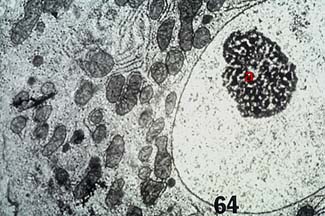
NUCLEUS
- Largest compartment in most cells.
- Control center. Contains chromosomes . If removed, cell works for a while, peters out
- not found in prokaryotes
- Bounded by nuclear membrane = double layered structure. Contains many nuclear pores , allow material to move in and out of nucleus.
- View nuclear pores
- Pores have octagonal "doors" made of protein; open and close on either side depending on specific signals.
- View diagram of nuclear pore structure
- Nucleus contains chromatin = DNA + proteins. Chromatin can condense into chromosomes during cell division. Contains DNA complexed to histone proteins =chromatin. During division, chromatin condenses by tight coiling into chromosomes, may be visible in light microscope. Machinery can handle large numbers of chromosomes, from 2 to over 1000. Very different from bacterial DNA separation (no spindle involved, no chromosome condensation, only 1 circular DNA molecule/cell (unless replicated prior to division).)
- View TEM of nucleus showing condensed chromatin ( heterochromatin ) and dispersed chromatin ( euchromatin )
- DNA remains in nucleus; information is exported in form of messenger RNA. mRNA is synthesized in nucleus, processed, and exported into cytoplasm to direct protein synthesis.
- Nucleolus = assembly plant for ribosomes. Ribosomal proteins are made in cytoplasm, must be transported back into nucleus. Ribosomal RNA made in nucleus. These two elements are integrated inside nucleolus to create ribosomal subunits. These are then exported out of nucleus through nuclear pores.
CYTOPLASM
- Fluid compartment. Filled with water, thousands of different cell molecules.Most metabolism occurs here (chemical transformations): food ---> energy,building blocks for growth.
- View TEM of a cell ; the clear zones inside the membrane are the cytoplasm (cytosol)
- Contains many ribosomes = sites of protein assembly.
RIBOSOMES
- Ribosome = assembly factory to make many PROTEINS in cytoplasm. Proteins direct most interesting cell processes.
- View ribosomes attached to endoplasmic reticulum membrane system (rER)
- Ribosome size measured in Svedberg (S) units; derived from sedimentation inultracentrifuge (used before electron microscopes were available)
- Prokaryotes: ribosomes made of 30S and 50S subunits, assemble into 70S ribosome
- Eukaryotes: ribosomes made of 40S and 60S subunits, assemble into 80S ribosome
- View schematic diagram of ribosome subunit structure
- In bacteria, ribosomes occupy 25% of cell volume, use 90% of cell energy. Less in many specialized eukaryotic cells, but still the dominant activity of almost all cells.
Endomembrane system
- System of membranes inside cell: parts able to move from one to another
- Endoplasmic Reticulum (ER): network of membranes inside cell.Includes both smooth and rough ER.
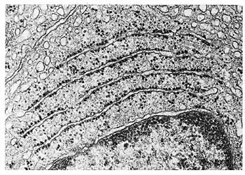
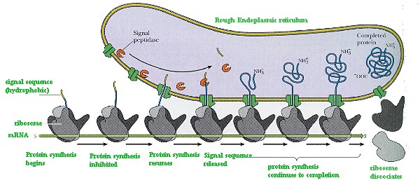
- rough ER (rER): most extensive part of endomembrane system. Synthesizes proteins for export or movement to different cell compartments (not to cytoplasm).
Signal hypothesis :certain mRNAs encode proteins designated for export. These carry a peptide signal at growing end, causes growing protein to move to ER ("docking"), insert peptide into membrane, translocate growing polypeptide chain across Er membrane. When protein synthesis is complete, polypeptide folds up inside ER,not in cytoplasm.- View diagram showing how signal hypothesis works
- Smooth ER (sER): synthesizes lipids, detoxifies drugs and poisons (in liver),
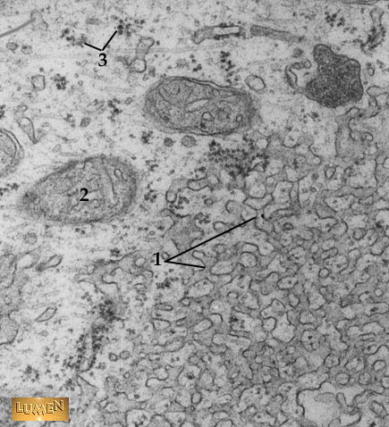
- View TEM of sER in a cell , including some free ribosomes (#3)
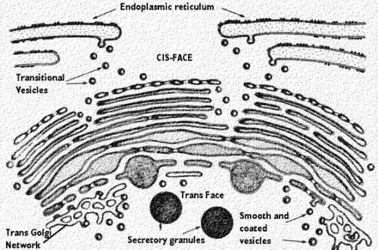
- Golgi body : functions as intracellular "post office" for sorting new proteins made on rER.
- View diagram showing Golgi organization
- Vesicles containing protein pinch off from ER,
- fuse with cis face of Golgi.
- Inside Golgi, oligosaccharide chains on proteins a remodified.
- Vesicles pinch off from trans face of Golgi, carry proteins to several possible destinations: export (out of cell), lysosomes, peroxisomes,cell membrane, etc.
- View animation showing movement from rER through endomembrane system to cell membrane (from The Biology Place)
- Lysosomes ( protected): compartments to break down old proteins, foreign materials, many wastes. Contain ~40 hydrolytic enzymes: lipases, proteases,nucleases, etc. Break down organic polymers of all types. "Suicide bags" if opened up on cell itself = apoptosis.
- View animation showing movement from rER through endomembrane system to lysosome (from The Biology Place)
- Vacuoles : large membrane compartments (contrasted with small membrane bags called vesicles ). Plant cells have especially large vacuole called the central vacuole, can occupy most of the volume of a plant cell. Stores pigments, wastes, water, poisons, and more.
CYTOPLASMIC ORGANELLES
- Organelles are not part of endomembrane system. All enclosed by double membrane system,contain DNA and ribosomes (70S), make some of their own proteins from their own genes. Divide by binary fission. Not autonomous, cannot grow or sustain life outside of cell.
- Mitochondria: diagram and EM Image ( protected)
- Centers for respiratory metabolism: Oxygen combined with chemicals to breakdown foods, generate cell energy. Contain outer and inner compartments, with many membranous cristae that "criss-cross" the internal space.
- Found in virtually every eukaryotic cell.
- Small structures similar to bacteria in some size.
- Chloroplasts: diagram and EM Image ( protected)
- Belong to group of plant organelles called plastids. Include chloroplasts(photosynthesis), amyloplasts (store starch), chromoplasts (store pigments).
- Chloroplasts = special organelles in plants. Trap light, convert energy to sugars (+ CO2, water). Contain stacks of thylakoids , where green pigmented chlorophyll is embedded in membrane to trap light.
Endosymbiont theory:
All organelles seem to share many properties with bacteria: contain 70S ribosomes (whereas rest of eukaryote cells contain 80S ribosomes), divide by binary fission, contain circular DNA without nucleus, etc. Lynn Margulis proposed endosymbiont hypothesis: that organelles derived from ancient colonization of large bacteria (became the eucaryotic cell) by smaller bacteria(became the mitochondria, chloroplast, etc.) Symbiosis = "living together".Eventually, organelles lost ability to exist as separate organisms, cannot be separated from cell. Recent evolutionary taxonomy by comparing ribosomal RNA shows that this idea has lots of merit. Mitochondrial and plastid ribosomes are very similar to current bacteria, very different from eukaryotes.CYTOSKELETON
- cell is not "just a bag in a bubble"; lots of internal fibers ---> internal skeleton. Not rigid; capable of being assembled, broken down in minutes. Allows cell movement, cell division, internal motion of compartments.
- Several different types of fibrous proteins make up cytoskeleton:
- Microtubules : one kind of fiber. Found in cytoplasm of all eukaryotes.Involved in many structures: cilia, flagella, spindle fibers that polymerize from centrioles during mitosis/meiosis. Made of tubulin protein; assembles into hollow tubules 25 nm diameter.
- Example 1: Cilia and flagella
- Used in locomotion
- 9 double rings of microtubules, 2 central microtubules.
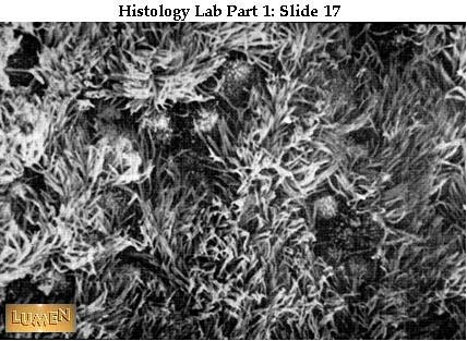
- View SEM of cilia on surface of epithelial tissue
- View fine structure of cilia ( protected)
- Two motor proteins allow motion along microtubules
View motor activity along microtubules
- Motor protein 1 -- Dynein:
- side arms allow one tubule to "walk up" along neighboring tubule, cause bending (powered by ATP).
- Causes motion from positive (+) end of the microtubule (where new tubulin is added to the microtubule) toward the minus (-) end of the microtubule.
- When coordinated, this causes rotating or whip-like motion of entire cilium or flagellum --->motion. move from the
- View animation of dynein pulling vesicle along a microtubule
- View cilia rotating as dynein causes sequential motion of microtubules
- Motor protein 2 -- Kinesin:
- Also powered by ATP, also allows protein to move along microtubule
- Causes motion from negative (-) end of the microtubule toward the positive (+) end of the microtubule (where new tubulin is added to the microtubule).
- pulls things toward outer reaches of cell
- Example: in nerve cells, kinesin pulls vesicles away from center towards nerve endings.
- View animation of kinesin pulling vesicle along a microtubule
- Example 2: Centrioles
- Microtubules usually grow out of structures called centrosomes
- In animal cells, centrosome = pair of centrioles, each has 9 sets of triplet microtubules in ring ("9" + "0")
- View centrosome organization ( protected)
- View centrosome with associated microtubules
- Microfilaments
- another kind of fiber. Found in cytoplasm of most eukaryotes. Same as actin protein. Involved in muscle contraction, cell support, pinching off of daughter cells after mitosis (in animals), cytoplasmic streaming (in plants).
- View TEM of microvilli ( protected)
- View animation showing "treadmilling" of actin -- subunits are added at one end, removed at the other, producing net migration of the filament in one direction.
- View movie showing cell movement (left panel) and assembly of actin (right panel) (1.7 Meg file)
- Intermediate filaments : a third kind of fiber. Made from keratin subunits. Not so quickly assembled and disassembled as microtubules or microfilaments. May be involved in resisting tension, reinforcing cell shape,fixing location of nucleus.
CELL WALLS
- Found in plants, fungi, bacteria -- not in animal cells
- Allow cells to survive in plain water, rigid structure (tree towering 150 feet high!)
- Thicker than cell membrane. Made from cellulose (plants and fungi), other polysaccharides (bacteria).
- View plant cell wall ( protected)
- Cells maintain contact by plasmodesmata -- thin cytoplasmic connections, lined by membrane, that pass across cell wall junctions.
EXTRACELLULAR MATRIX (ECM)
- Animal cells don't have walls, but do have ECM = meshwork of macromolecules outside plasma membrane. Consists mainly of glycoproteins(proteins with oligosaccharide chains), especially collagen.
- Some cells attached directly to ECM by bonding to collagen or fibronectin.
- View diagram of ECM ( protected)
INTERCELLULAR JUNCTIONS (see text for examples)
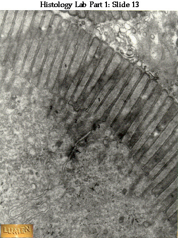
- View TEM of epithelial cell junction showing intercellular junctions. (1) "brush border" in intestinal cells; (2) microfilaments; (3) tight junctions; (4) desmosome
- Gap junctions (found in animals): formed by two connecting protein rings embedded in cell membrane of adjacent cells. Allows passage of water, small solutes, but not macromolecules (proteins, nucleic acids).
View animation of gap junctions (at The Biology Place)- Tight junctions (found in animals): specialized "belts" that bind two cells tightly to each other, prevent fluid from leaking into intracellular space.
View animation of tight junctions (at The Biology Place)- Desmosomes (found in animals): intercellular "rivets" that create tight bonds between cells, but allow fluids to pass through intracellular spaces.
View animation of desmosomes (at The Biology Place)- Plasmodesmata (found in plants): channels connecting cells; allow free passage of water and small solutes, but not macromolecules (proteins, nucleic acids).
View figure showing plasmodesmata (at The Biology Place)
Take a self-quiz on this lecture
Return to Lecture Index
Return to Biology 107 Index page